Alveoli Drawing
Alveoli Drawing - The alveoli are only one cell thick, allowing the relat. This article discusses the structure and function of the alveoli. This video helps you to draw science diagra. Web each alveolus is a microscopic sac lined with a single layer of flattened epithelial cells. Structure of alveoli in english. Let's look at the structure of alveoli, which makes them so suitable for the exchange of gases. The lining single layer of squamous cells (pneumocytes) can be seen peaking through the vessels. There are tiny blood vessels ( capillaries) surrounding each alveolus, and it is through these capillaries that oxygen and carbon dioxide are exchanged. Web this video explains how to draw alveolus diagram : The elastic fibers allow the alveoli to stretch as they are filled with air during inhalation.
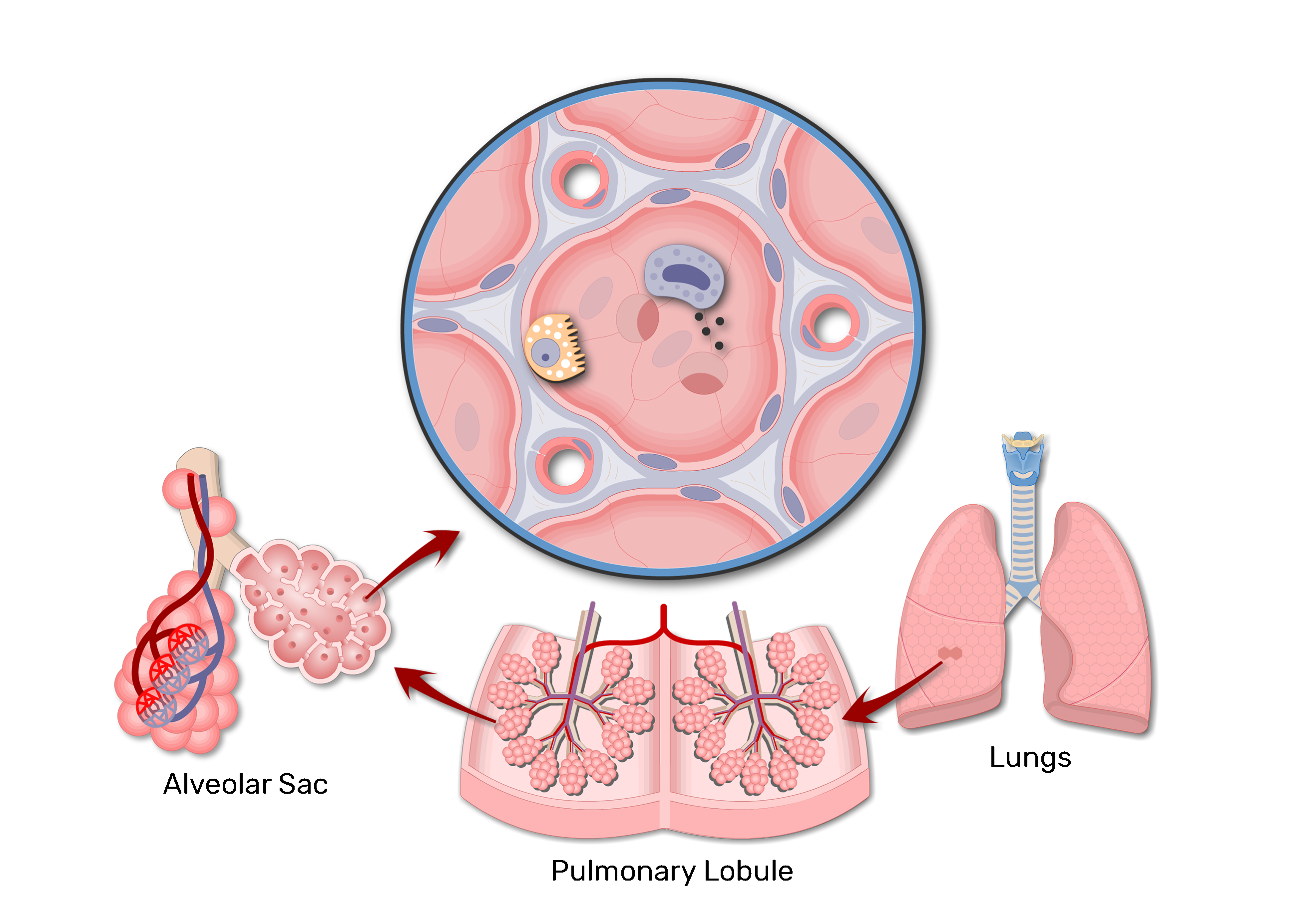
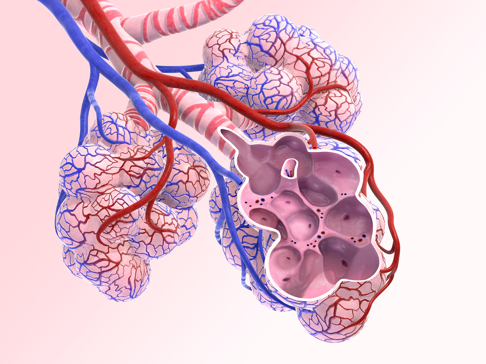
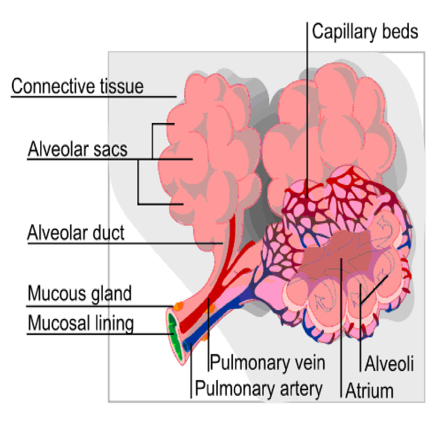
This article discusses the structure and function of the alveoli. Web this drawing demonstrates the open mouth view of the alveolus, which is surrounded by its capillary network. The lining single layer of squamous cells (pneumocytes) can be seen peaking through the vessels. Drawing shows the right lung with the upper, middle, and lower lobes, the left lung with the upper and lower lobes, and the trachea, bronchi, lymph nodes, and diaphragm. The respiratory bronchioles inside a secondary pulmonary lobule gives rise to two or more alveolar. View drawing of the alveoli videos. Structure of alveoli in english. This video explains how to draw alveolus diagram : The alveoli are located in the respiratory zone of the lungs, at the distal termination of the alveolar ducts. Web the alveoli consist of an epithelial layer and extracellular matrix surrounded by capillaries.
The lining single layer of squamous cells (pneumocytes) can be seen peaking through the vessels. A diagram of the pulmonary alveolus. The respiratory bronchioles inside a secondary pulmonary lobule gives rise to two or more alveolar. The alveoli are located in the respiratory zone of the lungs, at the distal termination of the alveolar ducts. The alveoli have a large surface area and receive oxygen from the blood that goes through an exchange of gases, allowing carbon dioxide to be removed. Web i practiced drawing the different parts like the trachea, bronchi, and alveoli in simple ways. This video explains how to draw alveolus diagram : There are as many as 700 million alveoli in each lungs, where they facilitate gaseous exchange of oxygen and carbon dioxide between inhaled air and the bloodstream. Drawing of the alveoli stock illustrations. Although they’re microscopic, alveoli are the workhorses of your respiratory.
Lung Alveolus Structure Lung Alveoli Anatomy GetBodySmart
Human internal organs set hand drawn vector. The alveoli are located in the respiratory zone of the lungs, at the distal termination of the alveolar ducts. Web this drawing demonstrates the open mouth view of the alveolus, which is surrounded by its capillary network. Each alveolus (singular) plays an important role in letting oxygen and carbon dioxide move into and.
:max_bytes(150000):strip_icc()/what-are-alveoli-2249043-01-94dfddd4dfe9488b8056d586824c7c36.png)
Alveoli Function, Lung Anatomy, and Causes of Damage
This video explains how to draw alveolus diagram : The lining single layer of squamous cells (pneumocytes) can be seen peaking through the vessels. View drawing of the alveoli videos. Drawing of an alveolus with blood capillary and gas exchange process, also shown type 1 and type 2 alveolar cells with secreted fluid surfactant. The alveoli move oxygen and carbon.

Alveoli anatomy. Human Respiratory Healthcare Illustrations
Structure of alveoli in english. One thing that really helped was finding guides that broke down the drawing into simple shapes. Drawing shows the right lung with the upper, middle, and lower lobes, the left lung with the upper and lower lobes, and the trachea, bronchi, lymph nodes, and diaphragm. Browse 190+ alveoli drawing stock photos and images available, or.
Alveoli crosssection A KYU Design
Web the alveoli of the lung are small air sacs that have extremely thin tissue walls. This video helps you to draw science diagra. Web alveoli are tiny balloon shaped structures and are the smallest passageway in the respiratory system. Drawing of an alveolus with blood capillary and gas exchange process, also shown type 1 and type 2 alveolar cells.

Draw Alveoli Graphical abstract Alveolus Scientific Illustration
3.1k views 1 year ago #biology #rahularts #science. Parts of respiratory system : Drawing of the alveoli stock illustrations. Let's look at the structure of alveoli, which makes them so suitable for the exchange of gases. Each alveolus (singular) plays an important role in letting oxygen and carbon dioxide move into and from the bloodstream during inhalation and exhalation [2,.

Alveoli, illustration Stock Image F028/9306 Science Photo Library
The alveoli contain some collagen and elastic fibres. Web learn the fine structure of the lungs. Drawing of the alveoli stock illustrations. The elastic fibers allow the alveoli to stretch as they are filled with air during inhalation. Web alveoli structure labeled diagram.

Lung alveoli anatomy and labeled diagram GetBodySmart
This video helps you to draw science diagra. Web this video explains how to draw alveolus diagram : Alveoli are tiny air sacs in your lungs that take up the oxygen you breathe in and keep your body going. Each alveolus (singular) plays an important role in letting oxygen and carbon dioxide move into and from the bloodstream during inhalation.
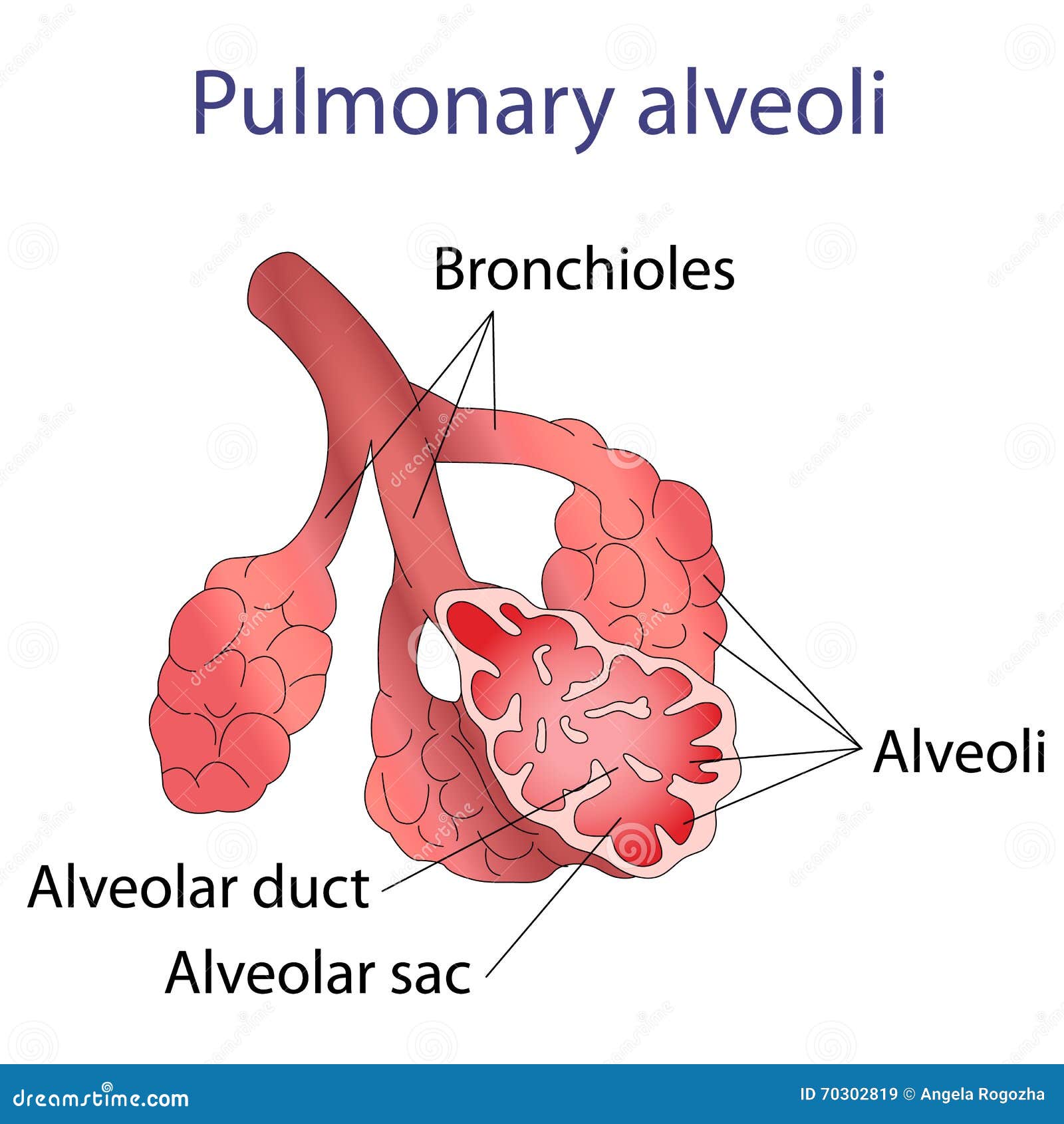
Illustration of Human Alveoli Structure Stock Vector Illustration of
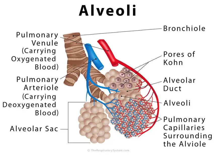
Structure of alveoli in english. The lining single layer of squamous cells (pneumocytes) can be seen peaking through the vessels. In some alveolar walls there are pores between alveoli called pores of kohn. 3.1k views 1 year ago #biology #rahularts #science. Web anatomy of the alveoli.
alveoli Critical Care Practitioner
By doing this, i could see how every part of the lungs fit together, like a puzzle. The respiratory bronchioles inside a secondary pulmonary lobule gives rise to two or more alveolar. The human lungs are a pair of large, spongy organs optimized for gas exchange between our blood and the air. The alveoli contain some collagen and elastic fibres..
Alveoli Definition, Location, Anatomy, Function, Diagrams
Parts of respiratory system : Web this drawing demonstrates the open mouth view of the alveolus, which is surrounded by its capillary network. View drawing of the alveoli videos. 3.1k views 1 year ago #biology #rahularts #science. Web learn the fine structure of the lungs.
Web This Video Explains How To Draw Alveolus Diagram :
This video helps you to draw science diagra. Web alveoli are tiny balloon shaped structures and are the smallest passageway in the respiratory system. Click to view large image. Drawing shows the right lung with the upper, middle, and lower lobes, the left lung with the upper and lower lobes, and the trachea, bronchi, lymph nodes, and diaphragm.
Web Anatomy Of The Alveoli.
Structure of alveoli in english. This video helps you to draw science diagra. The elastic fibers allow the alveoli to stretch as they are filled with air during inhalation. Structure of alveoli in english.
( 1 Vote) Video Transcript.
Each alveolus (singular) plays an important role in letting oxygen and carbon dioxide move into and from the bloodstream during inhalation and exhalation [2, 3]. The alveoli move oxygen and carbon dioxide (co 2) molecules into and out of your bloodstream. This article discusses the structure and function of the alveoli. In some alveolar walls there are pores between alveoli called pores of kohn.
What Age Is This For.
There are as many as 700 million alveoli in each lungs, where they facilitate gaseous exchange of oxygen and carbon dioxide between inhaled air and the bloodstream. 3.1k views 1 year ago #biology #rahularts #science. Parts of respiratory system : Browse 190+ alveoli drawing stock photos and images available, or start a new search to explore more stock photos and images.