Anatomical Drawing Of Lungs
Anatomical Drawing Of Lungs - Web human lungs drawing provides the artist with a knowledge of the human anatomy, specifically concerning the components of the body, responsible for breathing. 1.7m views 4 years ago biology drawing (easy). Web how to draw lungs. Web anatomy of the lungs. The current surge of 3d imaging analysis shows that the field is growing, with the technology continuing to improve. The right lung is situated on the right side of the heart and the mediastinum, while the left lung is. Web this article will discuss the anatomical basis of breathing and will describe the anatomical components that move every 5 seconds to keep you alive. Right and left lung are separated by the mediastinum. These are lined with simple squamosal epithelial. Web anatomy of the lungs.
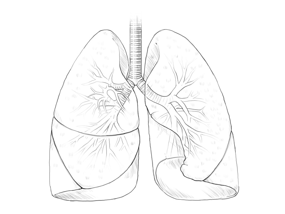
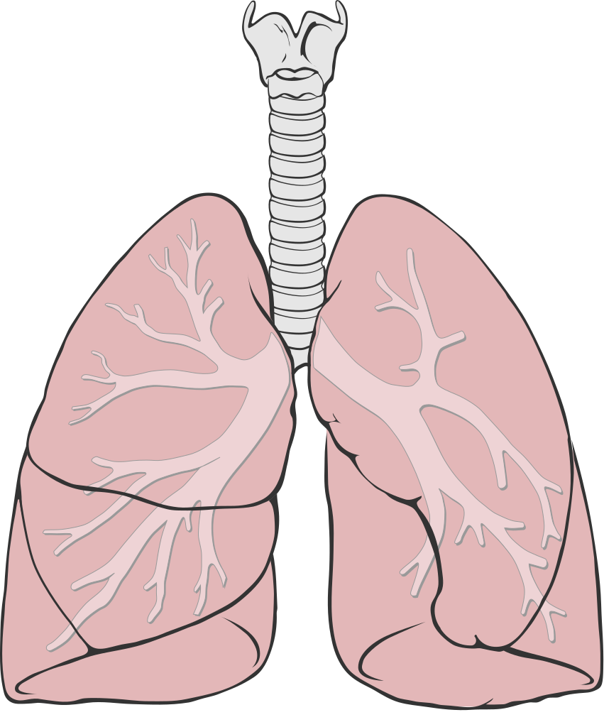
Web the lungs are roughly cone shaped, with an apex, base, three surfaces and three borders. They are a part of the respiratory system, which also includes the nose, nasal sinuses, mouth, pharynx, larynx, and trachea. Updated on august 16, 2023. By following the simple steps, you too can easily draw a perfect lungs. Web drawing and labeling the lungs can deepen your understanding of human anatomy. Jointly, the lungs inhabit most of the intrathoracic space. Unlike other epithelial tissue, the basement membrane of the tissue is connected to other squamosal epithelial cells; Start with the trachea, branching out into two main bronchi, and sketch the lobes. 561k views 3 years ago drawing and painting videos for kids. Web the lungs are the largest and main organs of the respiratory system.
These are lined with simple squamosal epithelial. February 5, 2024 | published on: 1.7m views 4 years ago biology drawing (easy). Either other alveoli or capillaries. Web anatomy of the lungs. Attached to the wall of the thoracic cavity, the parietal pleura forms the outer layer of the membrane. Web the lungs are the largest and main organs of the respiratory system. The visceral pleura forms the inner layer of the membrane covering the outside surface of the lungs. Web gross anatomy of lungs. Lungs diagram in human body.

How to Draw Lungs Really Easy Drawing Tutorial
Image information and view/download options. February 5, 2024 | published on: Updated on august 16, 2023. Acquire specific knowledge about the lungs that enables you to reason anatomically and solve anatomical and clinical. Web anatomy of the lungs.

How to Draw Lungs Really Easy Drawing Tutorial
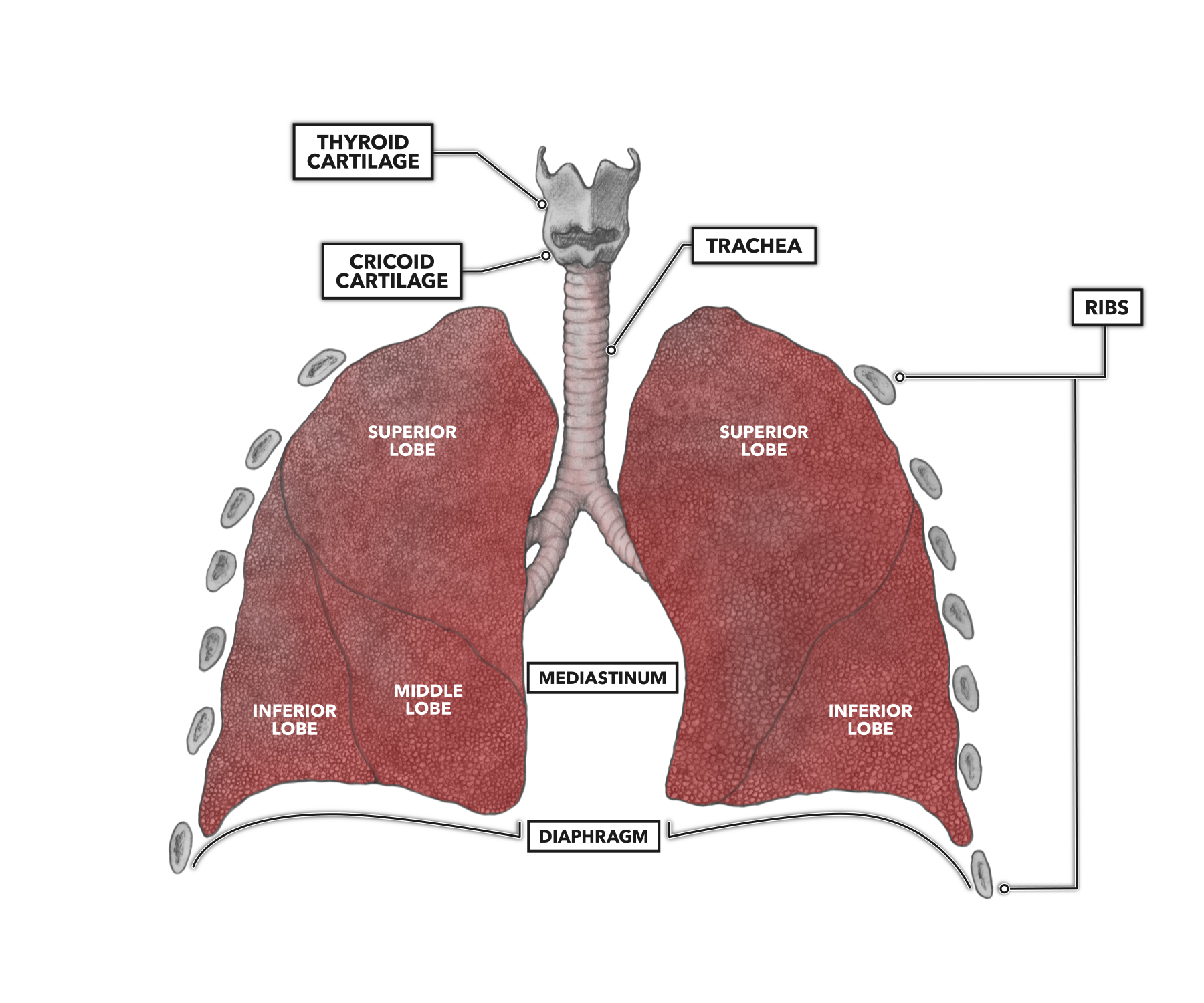
February 5, 2024 | published on: A spongy organ that moves oxygen through the bloodstream. It projects upwards, above the level of the 1st rib and into the floor of the neck. Web outline the anatomy of the blood supply to the lungs. Right and left lung are separated by the mediastinum.
Lungs Drawing at GetDrawings Free download
Either other alveoli or capillaries. They are a part of the respiratory system, which also includes the nose, nasal sinuses, mouth, pharynx, larynx, and trachea. Lungs are a pair of respiratory organs situated in a thoracic cavity. Web human lungs drawing provides the artist with a knowledge of the human anatomy, specifically concerning the components of the body, responsible for.

DRAWING OF HUMAN LUNGSDrawing and labelling of human lungs. Easy step
Hi everyone, in this video i show you how to draw the lungs step by step. These are lined with simple squamosal epithelial. Unlike other epithelial tissue, the basement membrane of the tissue is connected to other squamosal epithelial cells; Right and left lung are separated by the mediastinum. Every person has one right and one left lung.

Sketch human lungs anatomical organ Royalty Free Vector
561k views 3 years ago drawing and painting videos for kids. Illustrate the usefulness of the organ knowledge organization template (kot) for retrieving and organizing information associated with a specific organ; They are located on both sides of the mediastinum in the thorax. The current surge of 3d imaging analysis shows that the field is growing, with the technology continuing.

Section of the Lungs Anatomy Drawings 1888 illustrazione royaltyfree
A spongy organ that moves oxygen through the bloodstream. Web gross anatomy of the lungs. By following the simple steps, you too can easily draw a perfect lungs. A major organ of the respiratory system, each lung houses structures of both the conducting and respiratory zones. Attached to the wall of the thoracic cavity, the parietal pleura forms the outer.
CrossFit Anatomy of the Lungs
Describe the pleura of the lungs and their function. Web anatomy of the chest and the lungs: Humans have a right and a left lung positioned in the chest cavity. Medically reviewed by scott sundick, md. Web the lungs are roughly cone shaped, with an apex, base, three surfaces and three borders.

Human Lungs in Detail. Anatomy. Eps10 Vector Stock Illustration. Hand
Web outline the anatomy of the blood supply to the lungs. By following the simple steps, you too can easily draw a perfect lungs. Web the lungs are the main part of your respiratory system. Labeling parts like the bronchioles and alveoli can add educational value, making it informative and precise. 561k views 3 years ago drawing and painting videos.
FileLungs diagram simple.svg Wikipedia
On the inferior surface, the lungs are bordered by the diaphragm. They are located on both sides of the mediastinum in the thorax. A spongy organ that moves oxygen through the bloodstream. Web outline the anatomy of the blood supply to the lungs. Acquire specific knowledge about the lungs that enables you to reason anatomically and solve anatomical and clinical.

Single continuous line art anatomical human lungs Vector Image
The current surge of 3d imaging analysis shows that the field is growing, with the technology continuing to improve. The right lung is situated on the right side of the heart and the mediastinum, while the left lung is. Web gross anatomy of the lungs. Get free printable coloring page of this drawing. The visceral pleura forms the inner layer.
This Thoracic And Pulmonary Anatomy Tool Is Especially Designed For Students Of Anatomy (Medical And Paramedical Studies).
Web anatomy of the chest and the lungs: Attached to the wall of the thoracic cavity, the parietal pleura forms the outer layer of the membrane. Describe the pleura of the lungs and their function. The main function of the lungs is to perform the exchange of oxygen and carbon dioxide with air from the atmosphere.
Updated On August 16, 2023.
561k views 3 years ago drawing and painting videos for kids. Jointly, the lungs inhabit most of the intrathoracic space. The visceral pleura forms the inner layer of the membrane covering the outside surface of the lungs. These are lined with simple squamosal epithelial.
The Right Lung Is Situated On The Right Side Of The Heart And The Mediastinum, While The Left Lung Is.
Web human lungs drawing provides the artist with a knowledge of the human anatomy, specifically concerning the components of the body, responsible for breathing. Illustrate the usefulness of the organ knowledge organization template (kot) for retrieving and organizing information associated with a specific organ; It projects upwards, above the level of the 1st rib and into the floor of the neck. Acquire specific knowledge about the lungs that enables you to reason anatomically and solve anatomical and clinical.
In This Instance, The Lung.
Web gross anatomy of lungs. Get free printable coloring page of this drawing. 1.7m views 4 years ago biology drawing (easy). Web anatomy of the lungs.