Anatomy Of The Heart Drawing
Anatomy Of The Heart Drawing - Using basic shapes to create the general silhouette of the figure. This exercise will help you to identify your weak spots, so you’ll know which heart structures you need to spend more time. Web function and anatomy of the heart made easy using labeled diagrams of cardiac structures and blood flow through the atria, ventricles, valves, aorta, pulmonary arteries veins, superior inferior vena cava, and chambers. Web welcome to virtual heart. Xxxl very detailed human heart. Start with the pulmonary veins. Web drawings of the surface anatomy of the normal heart, anterior and posterior, with english labels. Web color human heart drawing. This interactive atlas of human heart anatomy is based on medical illustrations and cadaver photography. Web muscle and tissue make up this powerhouse organ.
Web the key to improving your drawings is to do your best and put your heart into your art. Web color human heart drawing. The left and right sides of the heart have different functions: Web the heart has three layers. It consists of four chambers, four valves, two main arteries (the coronary arteries), and the conduction system. Perfect the pencil drawing before moving on to the pen. Web cardiovascular system the cardiovascular system consists of the heart, blood vessels, and the approximately 5 liters of blood that the blood vessels transport. Simplifying forms for poses and gestures. Anatomical heart drawing stock photos are available in a variety of sizes and formats to fit your needs. Web his predecessor informed him the anatomy lab did a moment of silence to honor the donors and occasionally wrote letters to be included with the donors when they were sent to be cremated at the end of the academic year.
This is the superior vena cava. Let’s start with the building blocks of the human figure: Find a piece of paper and something to draw with. It also has several margins: The left and right sides of the heart have different functions: And cooking delicious healthy food. Web function and anatomy of the heart made easy using labeled diagrams of cardiac structures and blood flow through the atria, ventricles, valves, aorta, pulmonary arteries veins, superior inferior vena cava, and chambers. Web muscle and tissue make up this powerhouse organ. It consists of four chambers, four valves, two main arteries (the coronary arteries), and the conduction system. Adding in the appropriate level of detail depending on the style of.

Sketch of human heart anatomy line and color on a checkered background
The user can show or hide the anatomical labels which provide a useful tool to create illustrations perfectly adapted for teaching. Includes an exercise, review worksheet, quiz, and model drawing of an anterior view (frontal section) of the heart in. This exercise will help you to identify your weak spots, so you’ll know which heart structures you need to spend.

How to Draw the Internal Structure of the Heart 13 Steps
Web in this lecture, dr mike shows the two best ways to draw and label the heart! Your heart contains four muscular sections ( chambers) that briefly hold blood before moving it. Web muscle and tissue make up this powerhouse organ. The heart has five surfaces: The left and right sides of the heart have different functions:

Schematic Diagram Of Human Heart
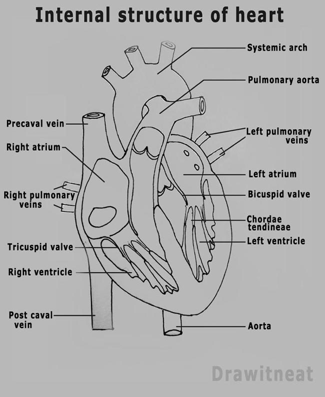
Web muscle and tissue make up this powerhouse organ. Web drawings of the surface anatomy of the normal heart, anterior and posterior, with english labels. Above this, draw a narrow vertical oval along the arch, and connect this to the atrium using a curved line. It may be a straight tube, as in spiders and annelid worms, or a somewhat.

How to Draw the Internal Structure of the Heart (with Pictures)
Electrical impulses make your heart beat, moving blood through these chambers. Heart anatomy drawing stock photos are available in a variety of sizes and formats to fit your needs. Three short quizzes regarding heart anatomy of each 10 questions, aimed at medical students. Above is a medical diagram of an anatomical heart drawing from an old nurses anatomy book. It.
Heart Anatomy Sketch at Explore collection of
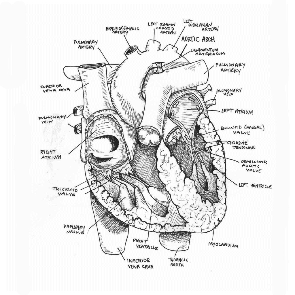
To find a good diagram, go to google images, and type in the internal structure of the human heart. Web muscle and tissue make up this powerhouse organ. Start with the pulmonary veins. Drawing from his previous work experience as the pathology/autopsy laboratory director at boston children’s. Next, draw the right atrium and the superior vena cava.
Anatomical Drawing Heart at GetDrawings Free download
Surgery colleagues to be innovative and creative in coming up with an individualized solution for each patient's unique anatomy and pathology. This interactive atlas of human heart anatomy is based on medical illustrations and cadaver photography. Above this, draw a narrow vertical oval along the arch, and connect this to the atrium using a curved line. Web with design your.

External Structure Of Heart Anatomy Diagram
Web heart drawing realistic anatomical beauty: Web anatomy of the human heart. Xxxl very detailed human heart. Simplifying forms for poses and gestures. Right, left, superior, and inferior:
DRAW IT NEAT How to draw human heart labeled
Web draw of a heart shape in the snow on the surface of a glass. This will make the pen drawing process much easier as we use the pencil marks for guidance. Web how to draw anatomy step by step. Build a character right from the start with the basics of human anatomy and proportion. Drawing internal anatomy of the.

Anatomy of the human heart
Base (posterior), diaphragmatic (inferior), sternocostal (anterior), and left and right pulmonary surfaces. Surgery colleagues to be innovative and creative in coming up with an individualized solution for each patient's unique anatomy and pathology. This exercise will help you to identify your weak spots, so you’ll know which heart structures you need to spend more time. This is the superior vena.
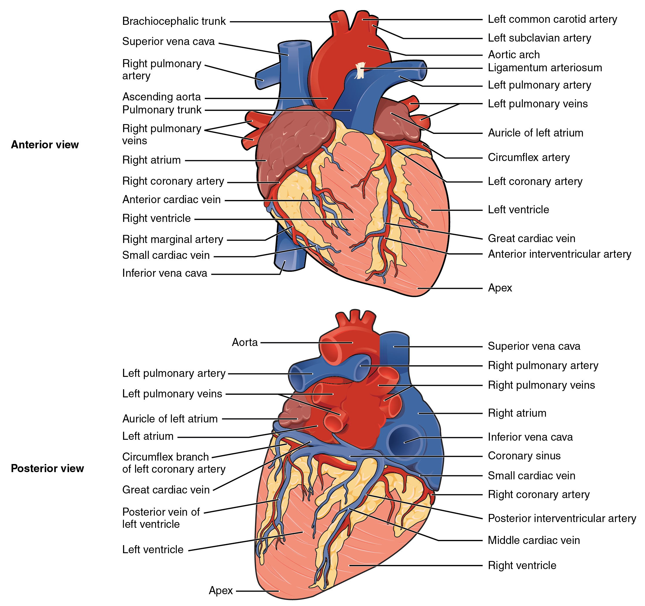
19.1 Heart Anatomy Anatomy and Physiology
Web in this lecture, dr mike shows the two best ways to draw and label the heart! Build a character right from the start with the basics of human anatomy and proportion. You can simplify the process of drawing human anatomy into three general steps: Pencil sketch of the human heart embark on a journey into the beauty of human.
It May Be A Straight Tube, As In Spiders And Annelid Worms, Or A Somewhat More Elaborate Structure With One Or More Receiving Chambers (Atria) And A Main Pumping Chamber (Ventricle), As In Mollusks.
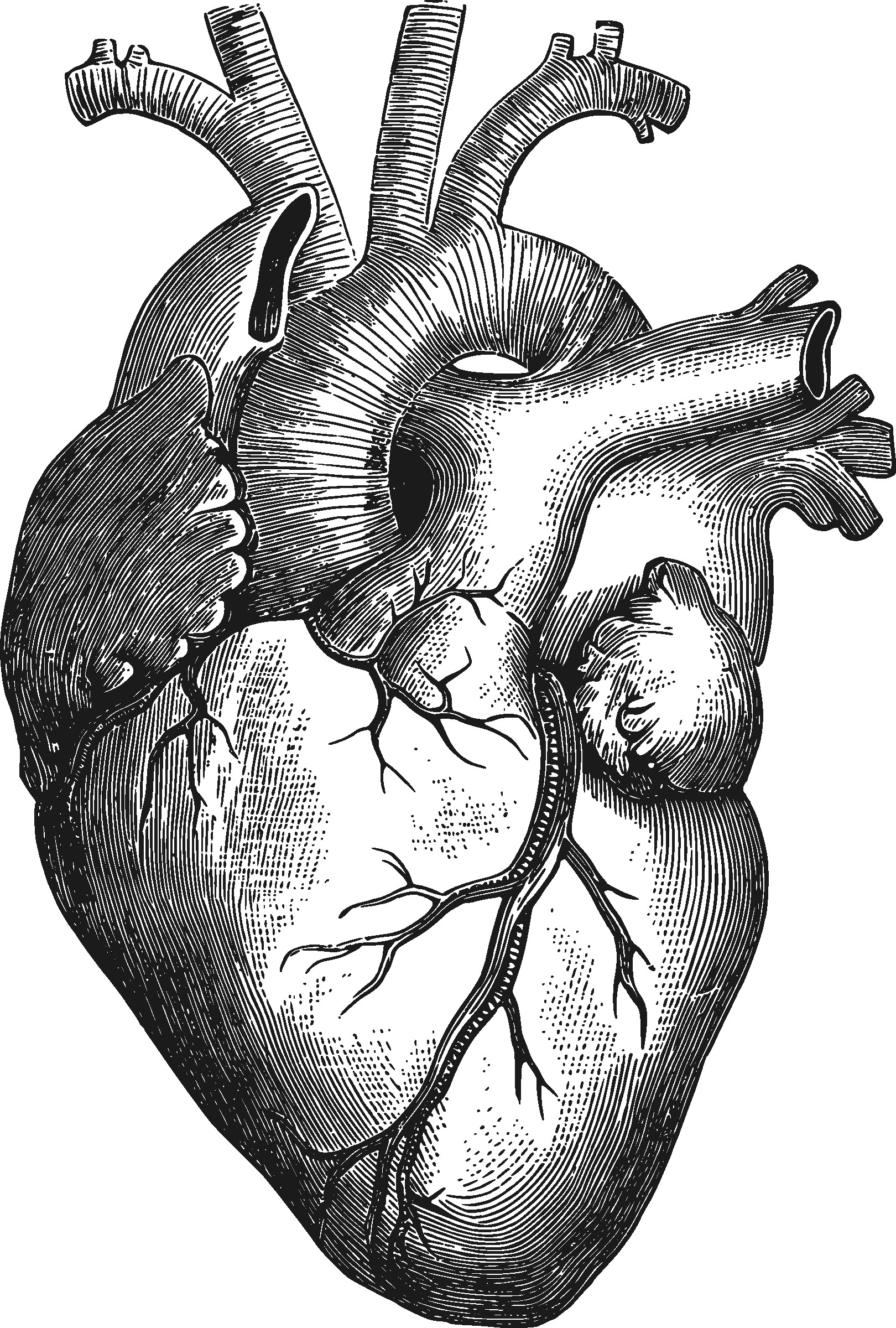
It is an anatomically correct heart drawing illustrating valves, veins, vessels, ventricles, chambers, etc. Pencil sketch of the human heart embark on a journey into the beauty of human anatomy with a mesmerizing pencil sketch of the human heart. Web his predecessor informed him the anatomy lab did a moment of silence to honor the donors and occasionally wrote letters to be included with the donors when they were sent to be cremated at the end of the academic year. The heart has five surfaces:
Web Cardiovascular System The Cardiovascular System Consists Of The Heart, Blood Vessels, And The Approximately 5 Liters Of Blood That The Blood Vessels Transport.
It is a full color heart illustration with blue and red sections along with black and white. Web the heart has three layers. Web muscle and tissue make up this powerhouse organ. Web heart drawing realistic anatomical beauty:
Create A “W” On Top Of The “J” Shape To Form The Narrow Tubes.
Heart anatomy drawing stock photos are available in a variety of sizes and formats to fit your needs. Using basic shapes to create the general silhouette of the figure. Build a character right from the start with the basics of human anatomy and proportion. From the openstax anatomy and physiology book.
Drawing Internal Anatomy Of The Heart.
Web welcome to virtual heart. This interactive atlas of human heart anatomy is based on medical illustrations and cadaver photography. Web minimally invasive heart surgery ; Electrical impulses make your heart beat, moving blood through these chambers.