Areolar Connective Tissue Drawing
Areolar Connective Tissue Drawing - Within the papillary layer are fibroblasts, a small number of. Web after the epithelium, i am posting videos on how to draw connective tissue. A few distinct cell types and densely packed fibers in a matrix characterize these tissues. In this photo of areolar connective tissue, nuclei of cells are stained but the cytoplasm is pale and not. Web brown adipose tissue is thermogenic, meaning that as it breaks down fats, it releases metabolic heat, rather than producing adenosine triphosphate (atp), a key molecule used in metabolism. Its cellular content is highly abundant and varied. Mesentery is bounded on their upper and lower surfaces by a simple squamous epithelium (the mesothelium) supported by a thin layer of collagen fibers. Collagen fibers are found in most supporting tissues and collagen is the most abundant protein in the body (wheaters). This is the well labelled diagram of structure of areolar tissue. Summary of the properties of the major types of connective tissue proper.
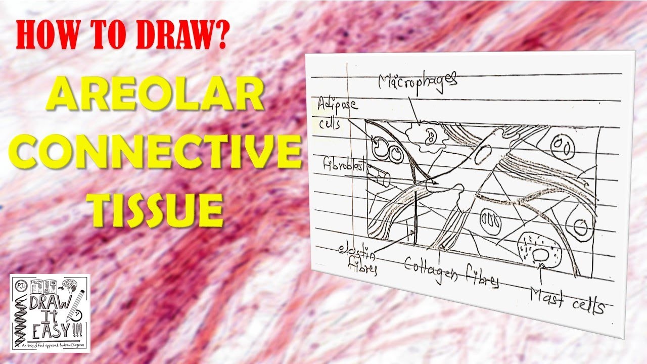
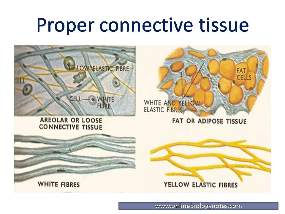
In drawing images of connective tissue proper preparations seen under the microscope, it is important to simplify the visuals. Loose connective tissue is composed of loosely woven collagen and elastic fibers. Web how to draw a diagram of areolar tissue in exam is the topic. Collagen fibers are found in most supporting tissues and collagen is the most abundant protein in the body (wheaters). Loose connective tissue, also called areolar connective tissue, has a sampling of all of the components of a. Web the areolar connective tissue is a type of connective tissue that is present throughout the human body. Areolar connective tissue is a soft tissue found in many areas including surrounding the blood vessels and nerves and forming the layer that flexibly attaches the skin to the muscles. The matrix has two components, fibers and ground substance. This loose fibrous tissue is widely distributed in the body where it provides strength, elasticity, and support to neighboring tissues. Web hello friends, this is my youtube channel and in this channel i used to share videos of different diagrams in easy way and step by step tutorials.
Web loose/areolar connective tissue figure 1. Fibroblasts are the cells that produce collagen and elastin that form the fibers found in connective tissue. In this photo of areolar connective tissue, nuclei of cells are stained but the cytoplasm is pale and not. Web after the epithelium, i am posting videos on how to draw connective tissue. Loose connective tissue is composed of loosely woven collagen and elastic fibers. This is a loose connective tissue that consists of fat cells with little extracellular matrix. Web brown adipose tissue is thermogenic, meaning that as it breaks down fats, it releases metabolic heat, rather than producing adenosine triphosphate (atp), a key molecule used in metabolism. A few distinct cell types and densely packed fibers in a matrix characterize these tissues. The ecm is composed of a moderate amount of ground substance and two main types of protein fibers: Many organs are suspended within the peritoneal cavity by mesenteries.
Areolar Connective Tissue Diagram
Its ground substance occupies more volume than the fibers do. Connective tissue preparations are often messy with a number of blotches and shapes irrelevant to the. Its cellular content is highly abundant and varied. Functions of the areolar connective tissue. The matrix has two components, fibers and ground substance.

Histology Drawing of Loose Areolar Tissue with explanation connective
This loose fibrous tissue is widely distributed in the body where it provides strength, elasticity, and support to neighboring tissues. Web this video will be very helpful for students to draw areolar tissue very easily thanks for watching and please subscribe to the channel for drawing sci. Areolar connective tissue is a soft tissue found in many areas including surrounding.
Areolar Connective Tissue Labeled Diagram
The matrix has two components, fibers and ground substance. Collagen fibers are found in most supporting tissues and collagen is the most abundant protein in the body (wheaters). Web brown adipose tissue is thermogenic, meaning that as it breaks down fats, it releases metabolic heat, rather than producing adenosine triphosphate (atp), a key molecule used in metabolism. Web the papillary.
chapter 4 connective tissues neuron stuff and other science stuff
This is a loose connective tissue that consists of fat cells with little extracellular matrix. Collagen fibers are found in most supporting tissues and collagen is the most abundant protein in the body (wheaters). The matrix has two components, fibers and ground substance. Web loose/areolar connective tissue figure 1. Web hello friends, this is my youtube channel and in this.

How to Draw Areolar Tissue Diagram Diagram of Areolar Tissue in
Web the papillary layer is made of loose, areolar connective tissue, which means the collagen and elastin fibers of this layer form a loose mesh. Loose connective tissue, also called areolar connective tissue, has a sampling of all of the components of a. Observe how the following are. Web #biology #class9 #class11 #areolarconnectivetissue #animaltissues #hscbiology #maharashtrastateboard2021 #biology2021 #biologydiagrams #icse #cbsethis.

How to Draw Areolar Tissue Diagram Step by Step //Areolar tissue
In bone, the matrix is rigid and described as calcified because of the deposited calcium salts. Web regular fibrous connective tissue is found in tendons (which connect muscles to bones) and ligaments (which connect bones to bones). Web loose/areolar connective tissue figure 1. Within the papillary layer are fibroblasts, a small number of. Fibrous connective tissue from the tendon has.
 Connective Tissue%2C 40X%2C Edited.jpg)
Areolar Connective Tissue Labeled Diagram
Its ground substance occupies more volume than the fibers do. Observe how the following are. Web the term areolar connective tissue means tissue with 'small open spaces' (areola) and refers to the appearance of small airy pockets between the network of cells and fibers. They usually stain pink and are the thickest fiber. Its cellular content is highly abundant and.

Areolar Connective Tissue Diagram Labeled
Loose connective tissue is composed of loosely woven collagen and elastic fibers. Web loose/areolar connective tissue figure 1. A few distinct cell types and densely packed fibers in a matrix characterize these tissues. Macrophages are present as well. Its ground substance occupies more volume than the fibers do.

Areolar Connective Tissue Slide Anatomy Physiology Flickr
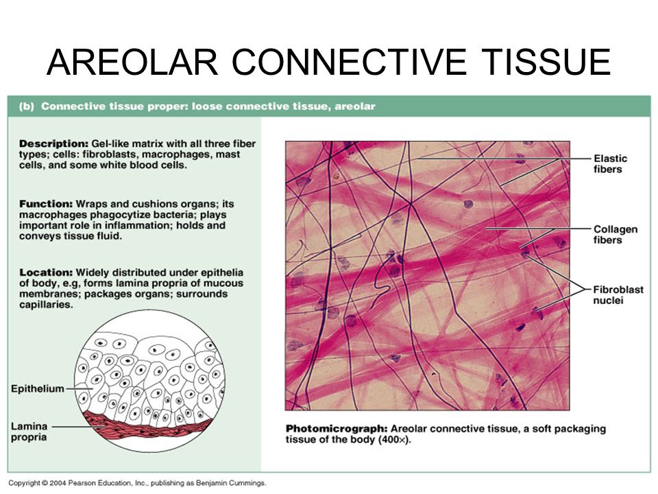
In this photo of areolar connective tissue, nuclei of cells are stained but the cytoplasm is pale and not. Collagen fibers are relatively wide and stain a light pink, while elastic fibers are thin and stain dark blue to black. Loose connective tissue (lct), also called areolar tissue, belongs to the category of connective tissue proper. It provides support and.

SOLUTION BIOL 1021 DU Mammalian & Epithelial Tissue Epidermis
Web regular fibrous connective tissue is found in tendons (which connect muscles to bones) and ligaments (which connect bones to bones). This loose fibrous tissue is widely distributed in the body where it provides strength, elasticity, and support to neighboring tissues. A few distinct cell types and densely packed fibers in a matrix characterize these tissues. This is the well.
In This Photo Of Areolar Connective Tissue, Nuclei Of Cells Are Stained But The Cytoplasm Is Pale And Not.
Many organs are suspended within the peritoneal cavity by mesenteries. The ecm is composed of a moderate amount of ground substance and two main types of protein fibers: Web the papillary layer is made of loose, areolar connective tissue, which means the collagen and elastin fibers of this layer form a loose mesh. Loose connective tissue is composed of loosely woven collagen and elastic fibers.
Web The Term Areolar Connective Tissue Means Tissue With 'Small Open Spaces' (Areola) And Refers To The Appearance Of Small Airy Pockets Between The Network Of Cells And Fibers.
Web the areolar connective tissue is a type of connective tissue that is present throughout the human body. The matrix has two components, fibers and ground substance. Web loose connective tissue, also called areolar connective tissue, has a sampling of all of the components of a connective tissue.as illustrated in figure 1, loose connective tissue has some fibroblasts; In drawing images of connective tissue proper preparations seen under the microscope, it is important to simplify the visuals.
Fibroblasts Are The Cells That Produce Collagen And Elastin That Form The Fibers Found In Connective Tissue.
Web regular fibrous connective tissue is found in tendons (which connect muscles to bones) and ligaments (which connect bones to bones). Loose connective tissue, also called areolar connective tissue, has a sampling of all of the components of a. Web loose/areolar connective tissue figure 1. This is the well labelled diagram of structure of areolar tissue.
Collagen Fibers Are Found In Most Supporting Tissues And Collagen Is The Most Abundant Protein In The Body (Wheaters).
This is a loose connective tissue that consists of fat cells with little extracellular matrix. Web supportive connective tissue —bone and cartilage—provide structure and strength to the body and protect soft tissues. Web how to draw a diagram of areolar tissue in exam is the topic. Its ground substance occupies more volume than the fibers do.