Bile Duct Drawing
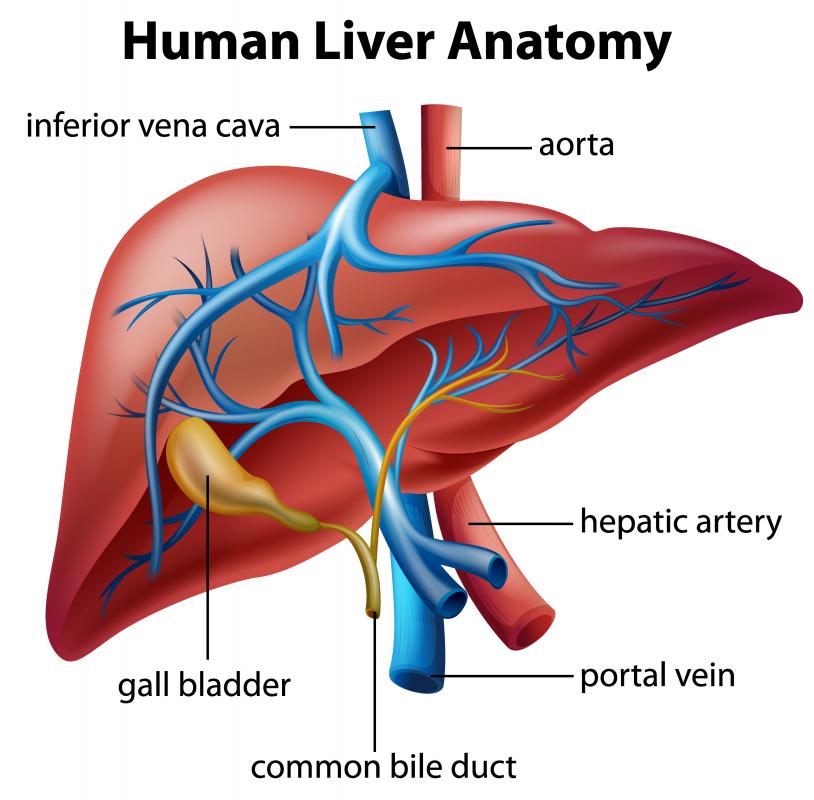
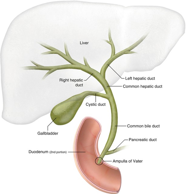
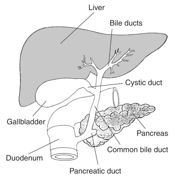
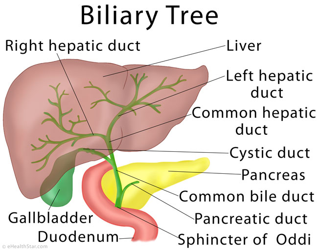
Bile Duct Drawing - Web the biliary system consists of the organs and ducts (bile ducts, gallbladder, and associated structures) that are involved in the production and transportation of bile. When the liver cells secrete bile, it is collected by a system of ducts that flow from the liver through the right and left. Their purpose is to carry bile between these organs. Web drawing of the biliary system, with the liver, gallbladder, duodenum, pancreatic duct, common bile duct, pancreas, cystic duct, and hepatic ducts labeled. Web drawing of the biliary system with the liver, biliary tree (bile ducts), common bile duct, gallbladder, pancreas, duodenal papilla, main pancreatic duct, and duodenum. All branches lead to the common bile duct, the main trunk of the biliary tree, which leads to your duodenum. Web your bile ducts collect bile where it’s created in your liver and carry it to the other organs in your biliary tract. The organs and bile ducts together form your biliary system. The biliary system is comprised of a system of these ducts, which flow from the liver to the gallbladder for storage and then into the small intestine (duodenum). You will find a drawing of the bile duct, which is the part of the body affected by bile duct cancer.
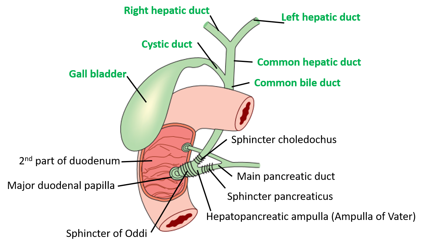
Gallstones are the most common cause. Once in the bulb the balloon can be filled slightly with water to maintain a stable position. Web drawing of the biliary system, with the liver, gallbladder, duodenum, pancreatic duct, common bile duct, pancreas, cystic duct, and hepatic ducts. Web the bile duct is visualized with a linear echoendoscope from either the duodenal bulb or the second portion of the duodenum. Web the common bile duct and the main pancreatic duct join before emptying their contents into the duodenum through the papillary orifice at the end of the duodenal papilla—a small, nipplelike structure that extends into the duodenum. Web the biliary system consists of the organs and ducts (bile ducts, gallbladder, and associated structures) that are involved in the production and transportation of bile. The bile duct is separated into three main parts: (0 indicates the absence of the symptom, while 1 indicates the presence of the symptom). It then runs in a groove near the. Web together with the bile duct, the hepatic portal vein and hepatic artery form the portal triad.
Web together with the bile duct, the hepatic portal vein and hepatic artery form the portal triad. The next section in this guide is risk factors and prevention. Web the common bile duct is formed by the junction of the cystic and hepatic ducts; Web draw a line down from each point to the probability axis to determine the likelihood of perforation in patients with choledochal cysts. Web drawing of the biliary system, with the liver, gallbladder, duodenum, pancreatic duct, common bile duct, pancreas, cystic duct, and hepatic ducts labeled. Their purpose is to carry bile between these organs. Bile ducts are tiny canals that connect some of the organs in your digestive system. Drawing shows the liver and the intrahepatic bile ducts, which include the right and left hepatic ducts. Treatment involves identifying what’s causing the blockage and removing it to prevent serious complications. The transportation of bile follows this sequence:
What is a Bile Duct? (with pictures)
Drawing shows the extrahepatic bile ducts, including the common hepatic duct (perihilar region) and the common bile duct (distal region). Web the biliary system consists of the organs and ducts (bile ducts, gallbladder, and associated structures) that are involved in the production and transportation of bile. Anatomy of the intrahepatic bile ducts; The biliary system is comprised of a system.

Biliary Tree Anatomy Bile duct, Human digestive system, Gallbladder
Web what is a bile duct? Their purpose is to carry bile between these organs. Web drawing of the biliary system with the liver, biliary tree (bile ducts), common bile duct, gallbladder, pancreas, duodenal papilla, main pancreatic duct, and duodenum. All branches lead to the common bile duct, the main trunk of the biliary tree, which leads to your duodenum..
Bile Duct Diagram
Web the anatomy of the intrahepatic bile ducts was typical in 63% of cases (n=188), showed triple confluence in 10% (n=29), anomalous drainage of the rpsd into the lhd in 11% (n=34), anomalous drainage of the rpsd into the common hepatic duct (chd) in 6% (n=19), anomalous drainage of the rpsd into the cystic duct in 2% (n=6),. Web the.
Bile Production, Secretion, Flow, Storage, Composition, pH, Function
Web drawing of the biliary system, with the liver, gallbladder, duodenum, pancreatic duct, common bile duct, pancreas, cystic duct, and hepatic ducts labeled. A bile duct drain, also called biliary drainage, can. It includes your liver, your gallbladder and your small intestine. Use the menu to see other pages. The transportation of bile follows this sequence:

(Color online) Anatomy of the biliary system. Download Scientific Diagram
This is a small, hollow tube that functions to transport bile. Drawing shows the extrahepatic bile ducts, including the common hepatic duct (perihilar region) and the common bile duct (distal region). Web the biliary system consists of the organs and ducts (bile ducts, gallbladder, and associated structures) that are involved in the production and transportation of bile. Web drawing of.
Biliary system with the liver, gallbladder, pancreas, duodenum, bile
Gallstones are the most common cause. Web drawing of the biliary system with the liver, biliary tree (bile ducts), common bile duct, gallbladder, pancreas, duodenal papilla, main pancreatic duct, and duodenum. Bile ducts are tiny canals that connect some of the organs in your digestive system. Web draw a line down from each point to the probability axis to determine.
Liver Anatomy, Location and Function eHealthStar
It includes your liver, your gallbladder and your small intestine. It then runs in a groove near the. These ducts amalgamate to form the common hepatic duct,. The next section in this guide is risk factors and prevention. All branches lead to the common bile duct, the main trunk of the biliary tree, which leads to your duodenum.

Pictures Of Biliary SystemHealthiack
Web schematic drawing shows (a) the typical intrahepatic bile ducts anatomy and (b) the anatomic variations; Web drawing of the biliary system, with the liver, gallbladder, duodenum, pancreatic duct, common bile duct, pancreas, cystic duct, and hepatic ducts. What is a bile duct drain? Web a bile duct drain is a procedure that involves opening up obstructions or treating holes.
Extrahepatic Biliary Apparatus Anatomy QA
Treatment involves identifying what’s causing the blockage and removing it to prevent serious complications. Web a bile duct drain is a procedure that involves opening up obstructions or treating holes in the biliary system. Web bile is initially secreted from hepatocytes and drains from both lobes of the liver via canaliculi, intralobular ducts and collecting ducts into the left and.

biliary tract Liver anatomy, Diagnostic medical sonography, Medical
Those structures supply blood to the sinusoids and the hepatocytes, subsequently draining into the central vein followed by the sublobular veins. Drainage of right posterior duct into right anterior duct on its lateral side, (c). Web drawing of the biliary system, with the liver, gallbladder, duodenum, pancreatic duct, common bile duct, pancreas, cystic duct, and hepatic ducts labeled. You will.
Web Drawing Of The Biliary System With The Liver, Biliary Tree (Bile Ducts), Common Bile Duct, Gallbladder, Pancreas, Duodenal Papilla, Main Pancreatic Duct, And Duodenum.
Drainage of right posterior duct into right anterior duct on its lateral side, (c). The transducer of the echoendoscope is advanced across the pylorus. It then runs in a groove near the. Also shown is the common hepatic duct, gallbladder, cystic duct, common bile duct, pancreas, ampulla of vater, and small intestine.
The Transportation Of Bile Follows This Sequence:
The organs and bile ducts together form your biliary system. Treatment involves identifying what’s causing the blockage and removing it to prevent serious complications. Those structures supply blood to the sinusoids and the hepatocytes, subsequently draining into the central vein followed by the sublobular veins. Web the common bile duct is formed by the junction of the cystic and hepatic ducts;
Bile Is A Digestive Product Made By The Liver.
Also shown are the liver, right and left hepatic ducts, gallbladder, cystic duct, pancreas, ampulla of vater, and small intestine. (0 indicates the absence of the symptom, while 1 indicates the presence of the symptom). All branches lead to the common bile duct, the main trunk of the biliary tree, which leads to your duodenum. Drawing shows the liver and the intrahepatic bile ducts, which include the right and left hepatic ducts.
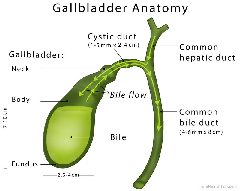
It Is About 7.5 Cm.
Web what is a bile duct? Bile ducts are tiny canals that connect some of the organs in your digestive system. Web the superiorbud and its associated stalk will become the gallbladder and cystic duct (respectively), while the inferior bud becomes the ventral component of the pancreas. Web the anatomy of the intrahepatic bile ducts was typical in 63% of cases (n=188), showed triple confluence in 10% (n=29), anomalous drainage of the rpsd into the lhd in 11% (n=34), anomalous drainage of the rpsd into the common hepatic duct (chd) in 6% (n=19), anomalous drainage of the rpsd into the cystic duct in 2% (n=6),.