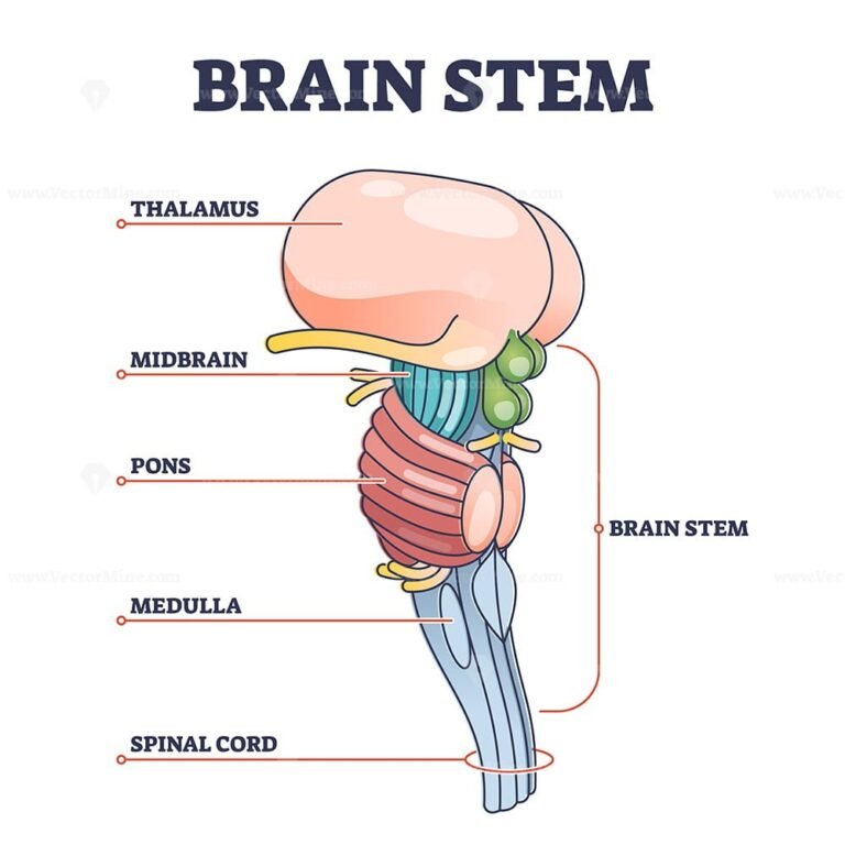
Brainstem Drawing
Brainstem Drawing - Superior to the ventricle are the inferior and superior colliculi. Web in this beautifully written paper, gates devises a system of approaching clinical brainstem neuroanatomy in a series of 4 simple rules. While studying the anatomy of the brainstem, it is helpful to remember the ‘rule of four’ of the. Localise a lesion on the brainstem from clinical features and. Web the medulla oblongata (medulla) is one of the three regions that make up the brainstem. The structure emerges from the ventral surface of the forebrain as a tapering cone that connects the brain to the spinal cord. Web the brainstem, positioned at the base of the posterior region of the brain, functions as the critical point of connection between the central and peripheral nervous systems. Web brainstem › drawing highlights. The brainstem (brain stem) is the distal part of the brain that is made up of the midbrain, pons, and medulla oblongata. Web master the medical sciences faster through our active learning approach to anatomy, biochemistry, biology, neuroanatomy, neuroscience, and physiology.
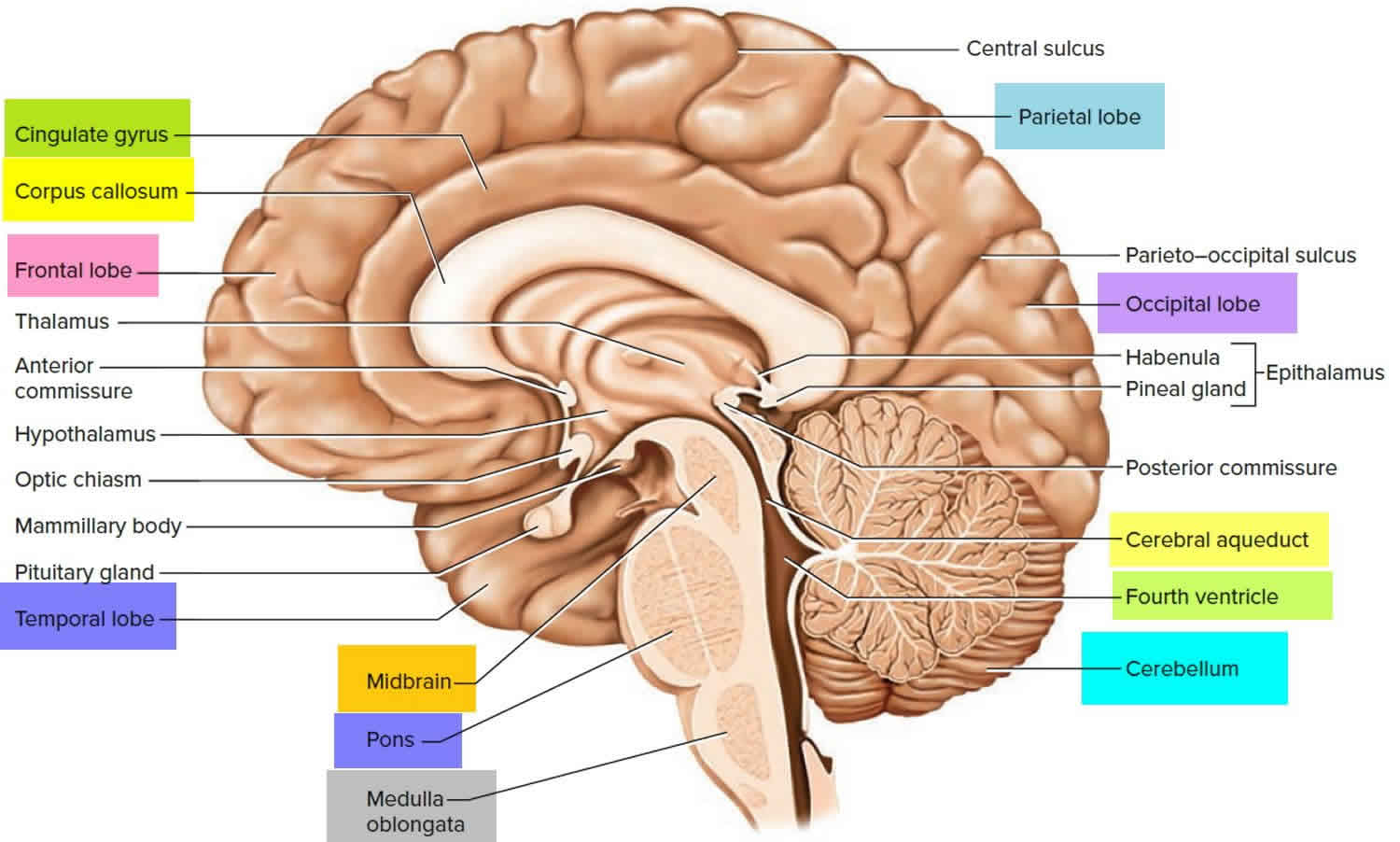
Web in this drawing they're showing sensory information coming in through cranial nerves to the brainstem, to different nuclei in different parts of the brainstem. Web in this beautifully written paper, gates devises a system of approaching clinical brainstem neuroanatomy in a series of 4 simple rules. Brainstem sections :how to draw. 2.1k views 1 year ago. Tell the various brainstem clinical syndromes. To begin, draw the cervical spinal cord. It's a stark contrast from what he learns within those walls. Web the medulla oblongata (medulla) is one of the three regions that make up the brainstem. While studying the anatomy of the brainstem, it is helpful to remember the ‘rule of four’ of the. The midbrain and hindbrain (composed of the pons and the medulla) are collectively referred to as the brain stem ( figure 11.3.1 11.3.
It is elegant, by explaining only that which can be detected by a basic neurological examination, and it is practical, by offering a system that can be easily remembered. Web the brainstem, positioned at the base of the posterior region of the brain, functions as the critical point of connection between the central and peripheral nervous systems. Web the brain stem. 152 part of the brain that interconnects the cerebrum and diencephalon with the spinal cord. Though small, the brainstem is an extremely important part of the brain, as the nerve connections from the motor and sensory systems of the cortex pass through it to communicate with the peripheral. Localise a lesion on the brainstem from clinical features and. This ventricle is rhomboid in shape, at its widest points it is bounded by the middle cerebellar peduncles. In the human brain, the brainstem is composed of the midbrain, the pons, and the medulla oblongata. The brainstem (brain stem) is the distal part of the brain that is made up of the midbrain, pons, and medulla oblongata. Begin segmenting the brainstem when the first slice containing the brainstem is visible.
/GettyImages-1092334754-fd0644493b3148288970e38fd26aead0.jpg)
Brainstem Anatomy, Function, and Treatment
Brainstem sections :how to draw. The medulla houses essential ascending and descending nerve tracts as well as brainstem nuclei. Web master the medical sciences faster through our active learning approach to anatomy, biochemistry, biology, neuroanatomy, neuroscience, and physiology. Ditki is the ideal resource for the flipped classroom: It is elegant, by explaining only that which can be detected by a.

Brain Anatomy and How the Brain Works Johns Hopkins Medicine
Web in this drawing they're showing sensory information coming in through cranial nerves to the brainstem, to different nuclei in different parts of the brainstem. Brainstem overview summarymajor nuclear groups • supplementary motor nuclei (eg,. Tell the various brainstem clinical syndromes. Brainstem sections :how to draw. It's a stark contrast from what he learns within those walls.
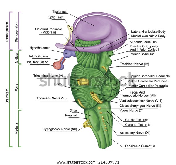
Brainstem Brain Stem Lateral View Posterior Stock Illustration 214509991
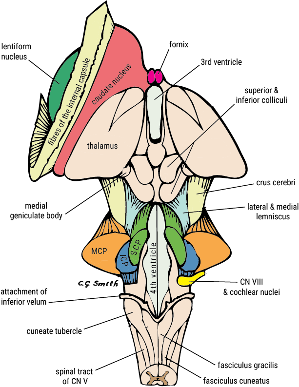
Learn the general anatomy of the brainstem by using a compressed composite of all three of its axial levels: Let's start with an anterior view of the brainstem, which is how we commonly study the brainstem in anatomy lab. Superior to the ventricle are the inferior and superior colliculi. Web the brain stem was represented on multiple angles, to show.
Brainstem Ventral View Posterior Part Brain Stock Illustration
Knowledge on ascending and descending nerve pathways, distribution of cranial nerve nuclei in. The midbrain and hindbrain (composed of the pons and the medulla) are collectively referred to as the brain stem ( figure 11.3.1 11.3. To begin, draw the cervical spinal cord. The structure emerges from the ventral surface of the forebrain as a tapering cone that connects the.
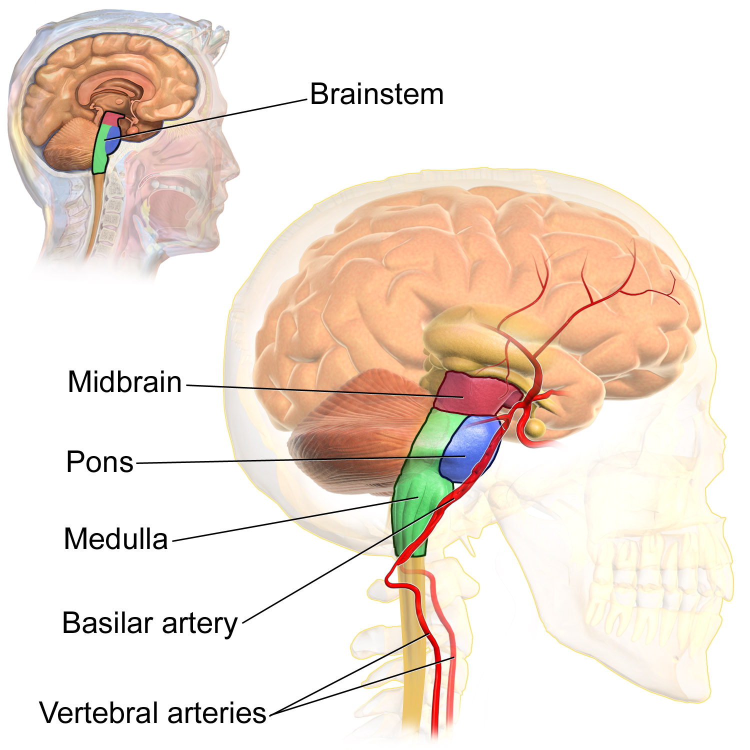
The brain stem Anatomy of the brain stem Physiology of the brain
Learn the general anatomy of the brainstem by using a compressed composite of all three of its axial levels: The midbrain and hindbrain (composed of the pons and the medulla) are collectively referred to as the brain stem ( figure 11.3.1 11.3. Superior to the ventricle are the inferior and superior colliculi. Web the medulla oblongata (medulla) is one of.
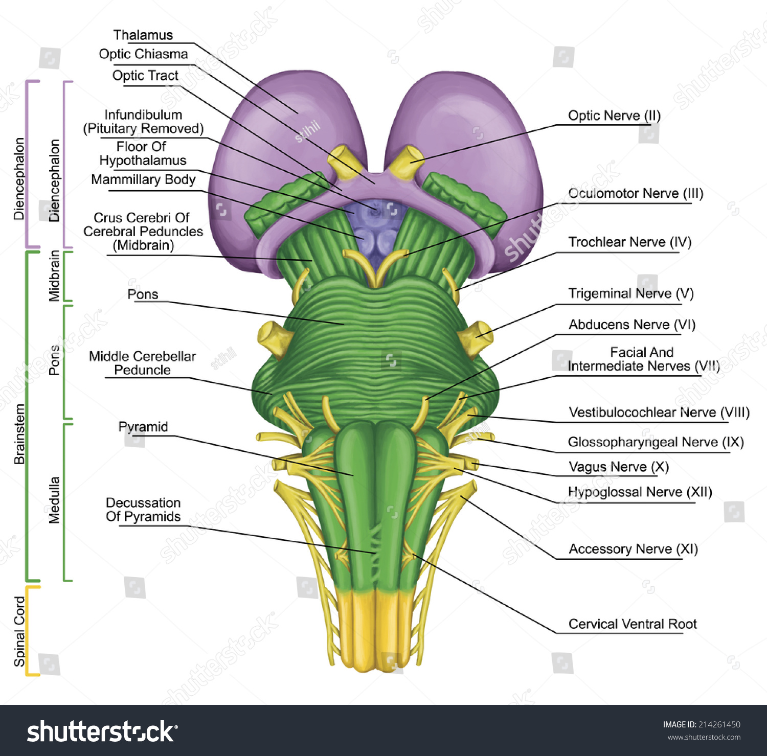
U.Br.Columbia Drawing Posterior view of brainstem English labels
Each of the three components has its own unique structure and function. It is elegant, by explaining only that which can be detected by a basic neurological examination, and it is practical, by offering a system that can be easily remembered. Web the brain stem was represented on multiple angles, to show the external structures of the midbrain, pons and.
Brain stem parts anatomical model in educational labeled outline
It is connected with other parts of the central nervous system, including the spinal cord, cerebellum, diencephalon, and cerebral hemispheres. Though small, the brainstem is an extremely important part of the brain, as the nerve connections from the motor and sensory systems of the cortex pass through it to communicate with the peripheral. In the human brain, the brainstem is.
Brain stem anatomy, function, brain stem stroke & brain stem tumor
The brainstem (brain stem) is the distal part of the brain that is made up of the midbrain, pons, and medulla oblongata. Learn the general anatomy of the brainstem by using a compressed composite of all three of its axial levels: Web brainstem › drawing highlights. Superior to the ventricle are the inferior and superior colliculi. Web the brainstem, positioned.

Brainstem Neurology Medbullets Step 1
Let's start with an anterior view of the brainstem, which is how we commonly study the brainstem in anatomy lab. The structure emerges from the ventral surface of the forebrain as a tapering cone that connects the brain to the spinal cord. Draw the anatomy of the midbrain, pons and medulla. New 3d rotate and zoom. The midbrain and hindbrain.

The brain stem and the cerebelleum Human Anatomy and Physiology Lab
The midbrain and hindbrain (composed of the pons and the medulla) are collectively referred to as the brain stem ( figure 11.3.1 11.3. To begin, draw the cervical spinal cord. Web in this drawing they're showing sensory information coming in through cranial nerves to the brainstem, to different nuclei in different parts of the brainstem. It is the most inferior.
Web The Brainstem, Positioned At The Base Of The Posterior Region Of The Brain, Functions As The Critical Point Of Connection Between The Central And Peripheral Nervous Systems.
Web the medulla oblongata (medulla) is one of the three regions that make up the brainstem. Web in vertebrate anatomy, the brainstem is the posterior part of the brain adjoining, and structurally continuous with, the spinal cord. Let's start with an anterior view of the brainstem, which is how we commonly study the brainstem in anatomy lab. There are twelve pairs of nerves that stem directly from the brainstem to control all senses that can be detected from the organs in the cranial region.
Tell The Various Brainstem Clinical Syndromes.
Superior to the ventricle are the inferior and superior colliculi. The brainstem (brain stem) is the distal part of the brain that is made up of the midbrain, pons, and medulla oblongata. It's a stark contrast from what he learns within those walls. Web master the medical sciences faster through our active learning approach to anatomy, biochemistry, biology, neuroanatomy, neuroscience, and physiology.
To Begin, Draw The Cervical Spinal Cord.
In the human brain, the brainstem is composed of the midbrain, the pons, and the medulla oblongata. The midbrain and hindbrain (composed of the pons and the medulla) are collectively referred to as the brain stem ( figure 11.3.1 11.3. This ventricle is rhomboid in shape, at its widest points it is bounded by the middle cerebellar peduncles. New 3d rotate and zoom.
Let's Start With An Anterior View Of The Brainstem, Which Is How We Commonly Study The Brainstem In Anatomy Lab.
Brainstem sections :how to draw. Localise a lesion on the brainstem from clinical features and. Knowledge on ascending and descending nerve pathways, distribution of cranial nerve nuclei in. Web this procedure exposes the pontomedullary junction as well as the floor of the fourth ventricle.