Cardiac Muscle Tissue Drawing
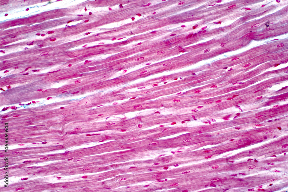
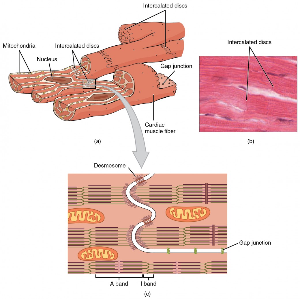
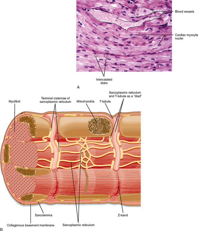
Cardiac Muscle Tissue Drawing - Intercalated discs are complex cell junctions between the ends of adjacent cardiac muscle fibers. Web circulatory system > heart anatomy > cardiac muscle tissue: Many more cardiac muscle cell nuclei are visible. It is one of three types of muscle in the body, along with skeletal and smooth muscle. The individual cardiac muscle cells are arranged in bundles that form a spiral pattern in the wall of the heart. Web 16/10/2023 17/12/2022 by sonnet poddar. Components of intercalated disc cannot be resolved with the light microscope. Cardiac muscle tissue contracts and. Web the cardiac muscle or the myocardium forms the musculature of the heart. The cardiac muscle under a microscope shows a short cylindrical fiber with a centrally placed oval nucleus.
Many more cardiac muscle cell nuclei are visible. Even though cardiac muscle has autorhythmicity, heart rate is modulated by the endocrine and nervous systems. You will find some unique features in cardiac muscle that will help you to differentiate it. How to draw a muscle. Similar to skeletal muscle, cardiac muscle is striated and organized into sarcomeres, possessing the same banding organization as skeletal muscle ( figure 10.21 ). These are striated and involuntary muscles that are supplied by autonomic nerve fibres. The cardiac muscle under a microscope shows a short cylindrical fiber with a centrally placed oval nucleus. It is the pen diagram of skeletal, smooth and cardiac muscle for class 10, 11 and 12. There are two major types of cardiac muscle cells: Lab 4 muscle and nervous tissue.
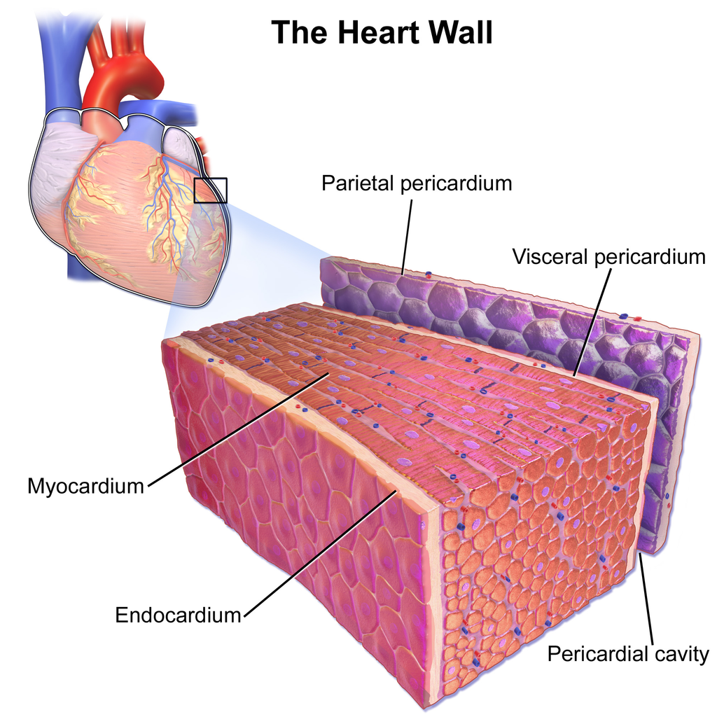
Web cardiac muscle cells are cylindrical cells whose ends branch and form junctions with other cardiac muscle cells. Its unique structural and functional characteristics enable the heart to perform its vital role of pumping blood throughout the body continuously and rhythmically. Cardiac muscle with intercalated discs. Cardiac muscle, or myocardium, is a specialized type of muscle found exclusively in the heart. Web cardiac muscle (or myocardium) makes up the thick middle layer of the heart. Web circulatory system > heart anatomy > cardiac muscle tissue: Web in this video i have shown the simplest way of drawing muscle drawing. In the connective tissue between cardiac. Components of intercalated disc cannot be resolved with the light microscope. It is capable of strong, continuous, and rhythmic contractions that are automatically generated.

How to draw " Cardiac Muscles" step by step in a very easy way Type
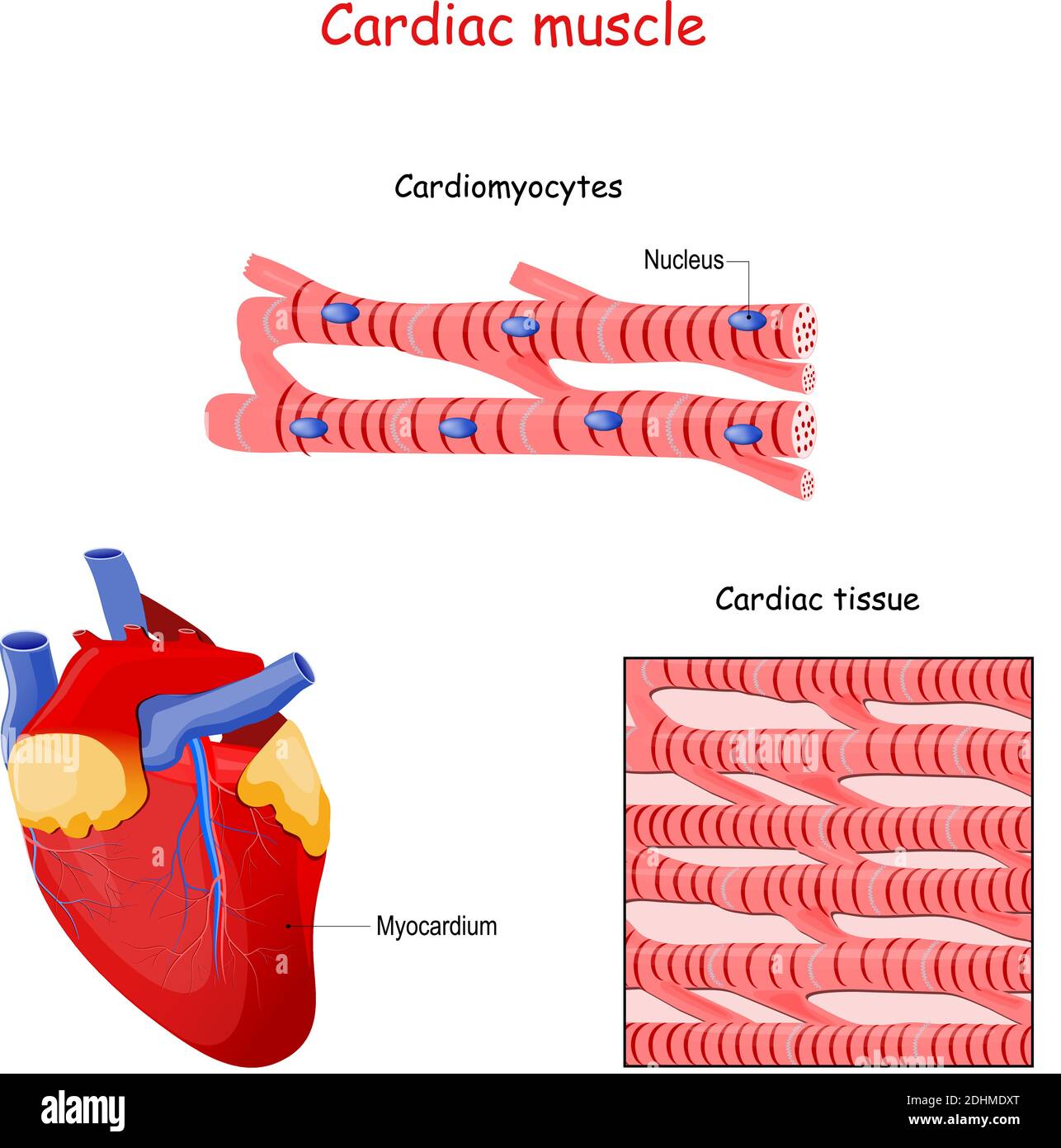
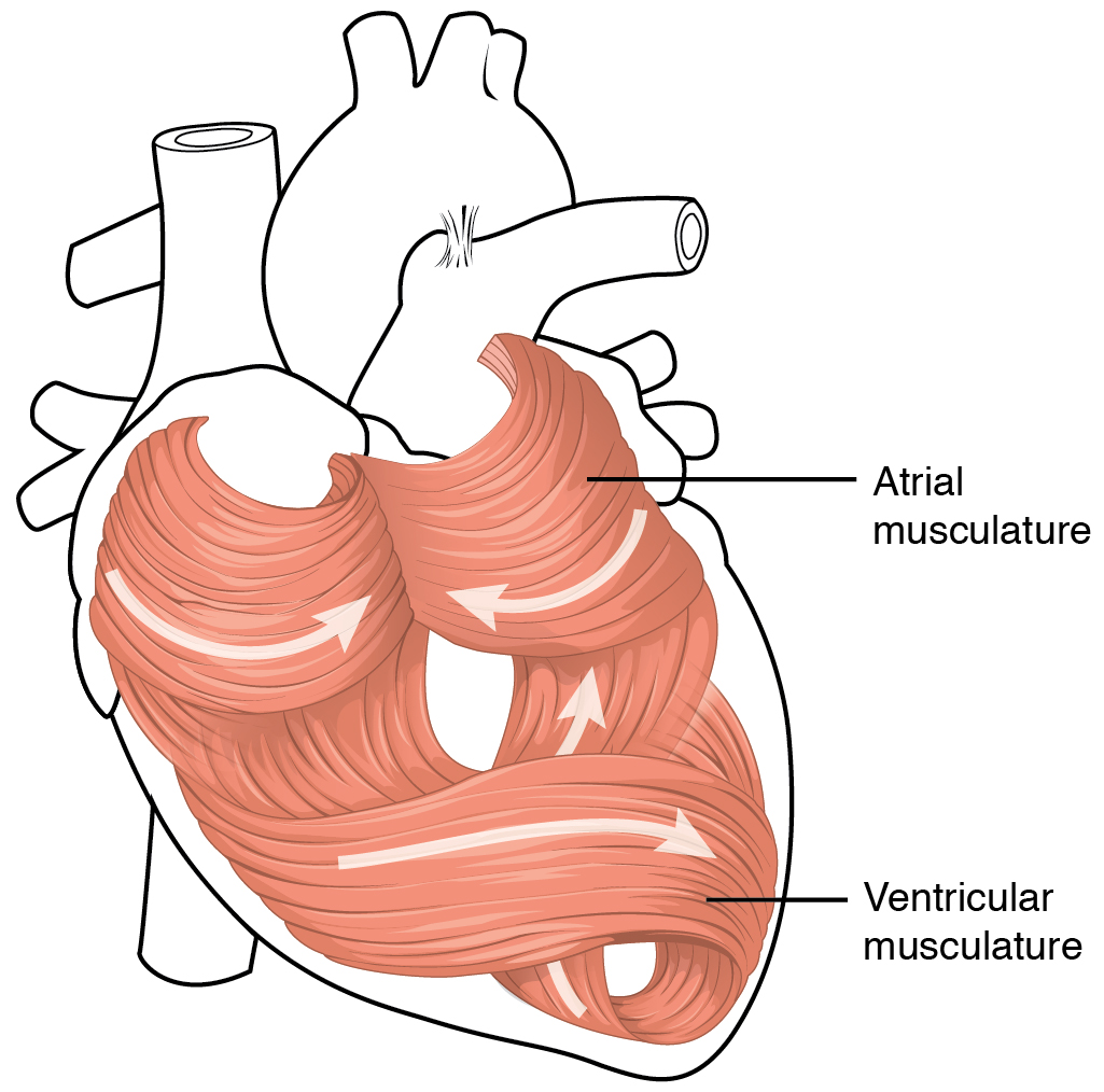
Describe intercalated discs and gap junctions. The individual cardiac muscle cells are arranged in bundles that form a spiral pattern in the wall of the heart. Highly coordinated contractions of cardiac muscle pump blood into the vessels of the circulatory system. Web table of contents. 5.9k views 2 years ago #class 9 science :.
Histology of human cardiac muscle under light microscope view for
Web draw cardiac muscle tissue diagram easily with this video. Web in this video i have shown the simplest way of drawing muscle drawing. You will find some unique features in cardiac muscle that will help you to differentiate it. Cardiac muscle tissue is only found in your heart. Cardiac muscle, in vertebrates, one of three major muscle types, found.
Cardiac Muscle and Electrical Activity Anatomy and Physiology II
Cardiac muscle tissue, or myocardium, is a type of muscle tissue that forms the heart. A cardiac muscle cell typically has one nucleus located near the center. Cardiac muscle, in vertebrates, one of three major muscle types, found only in the heart. Web 16/10/2023 17/12/2022 by sonnet poddar. Cross section of cardiac muscle fibers.

Simple histology diagram of Cardiac Tissue/ Muscle Longitudinal Section
Cardiac muscle tissue contracts and. Cardiac muscle, or myocardium, is a specialized type of muscle found exclusively in the heart. Web 16/10/2023 17/12/2022 by sonnet poddar. The individual cardiac muscle cells are arranged in bundles that form a spiral pattern in the wall of the heart. Many more cardiac muscle cell nuclei are visible.
Which is the cardiac muscle layer of the heart? Socratic
Similar to skeletal muscle, cardiac muscle is striated and organized into sarcomeres, possessing the same banding organization as skeletal muscle ( figure 10.21 ). The cardiac muscle under a microscope shows a short cylindrical fiber with a centrally placed oval nucleus. You will find some unique features in cardiac muscle that will help you to differentiate it. It is one.
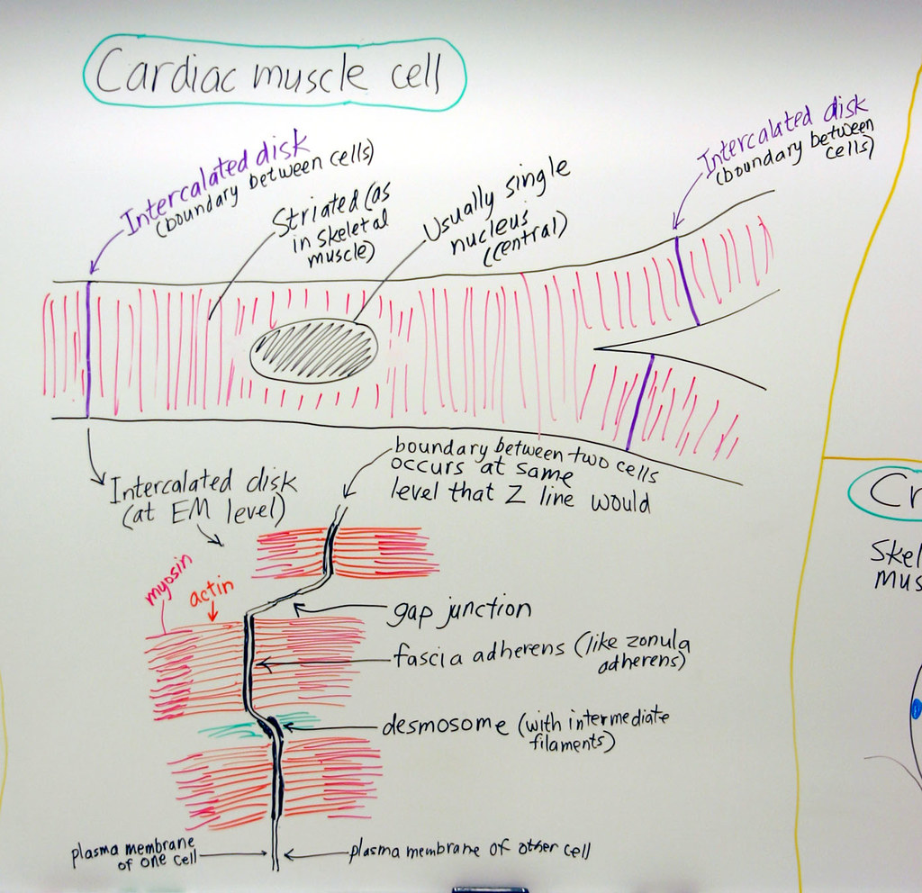
Muscle Cardiac Muscle Cell A hand drawn sketch by Dr. Chr… Flickr
Describe intercalated discs and gap junctions. Web 16/10/2023 17/12/2022 by sonnet poddar. Web draw cardiac muscle tissue diagram easily with this video. The individual cardiac muscle cells are arranged in bundles that form a spiral pattern in the wall of the heart. Let's learn more about the cardiac muscle with the help of a diagram.
Cardiac Muscle Structure
Cardiac muscle, in vertebrates, one of three major muscle types, found only in the heart. In the connective tissue between cardiac. A cardiac muscle cell typically has one nucleus located near the center. Let's learn more about the cardiac muscle with the help of a diagram. The cardiac muscle under a microscope shows a short cylindrical fiber with a centrally.
Cardiac Muscle Tissue Labeled Diagram
Highly coordinated contractions of cardiac muscle pump blood into the vessels of the circulatory system. Myocardial contractile cells and myocardial conducting cells. Web cardiac muscle cells are cylindrical cells whose ends branch and form junctions with other cardiac muscle cells. The cardiac muscle under a microscope shows a short cylindrical fiber with a centrally placed oval nucleus. There are two.
17.2 Heart Anatomy Medicine LibreTexts
Cardiac muscle, in vertebrates, one of three major muscle types, found only in the heart. Web 16/10/2023 17/12/2022 by sonnet poddar. How to draw a muscle. Web in this video i have shown the simplest way of drawing muscle drawing. Web table of contents.
12.3 Types of Muscle Tissue Human Biology
Similar to skeletal muscle, cardiac muscle is striated and organized into sarcomeres, possessing the same banding organization as skeletal muscle ( figure 10.21 ). Highly coordinated contractions of cardiac muscle pump blood into the vessels of the circulatory system. It is the pen diagram of skeletal, smooth and cardiac muscle for class 10, 11 and 12. The individual cardiac muscle.
It Is One Of Three Types Of Muscle In The Body, Along With Skeletal And Smooth Muscle.
Web cardiac muscle tissue is only found in the heart. Cardiac muscle, in vertebrates, one of three major muscle types, found only in the heart. Myocardial contractile cells and myocardial conducting cells. This feature, however, also distinguishes it from smooth muscle, the third muscle type.
These Inner And Outer Layers Of The Heart, Respectively, Surround The Cardiac Muscle Tissue And Separate It From The Blood And.
Web the cardiac muscle or the myocardium forms the musculature of the heart. Even though cardiac muscle has autorhythmicity, heart rate is modulated by the endocrine and nervous systems. Highly coordinated contractions of cardiac muscle pump blood into the vessels of the circulatory system. Web draw cardiac muscle tissue diagram easily with this video.
In The Connective Tissue Between Cardiac.
Describe intercalated discs and gap junctions. Intercalated discs are complex cell junctions between the ends of adjacent cardiac muscle fibers. Web circulatory system > heart anatomy > cardiac muscle tissue: Cardiac muscle, also known as heart muscle, is the layer of muscle tissue which lies between the endocardium and epicardium.
Cardiac Muscle Tissue, Or Myocardium, Is A Type Of Muscle Tissue That Forms The Heart.
Components of intercalated disc cannot be resolved with the light microscope. Web cardiac muscle tissue, also known as myocardium, is a structurally and functionally unique subtype of muscle tissue located in the heart, that actually has characteristics from both skeletal and muscle tissues. These are striated and involuntary muscles that are supplied by autonomic nerve fibres. A cardiac muscle cell typically has one nucleus located near the center.