Compound Microscope Drawing
Compound Microscope Drawing - Web microscope parts and functions with labeled diagram and functions how does a compound microscope work?. It is another type of optical microscope (the first one being a ‘simple microscope’). The part that is looked through at the top of the compound microscope. This stands by resting on the base and supports the stage. Web learn to draw compound microscope diagram (final image at least distance of distinct vision d) within 3 mins.background score credit: In contrast, “simple microscopes” have only one convex lens and function more like glass magnifiers. You and say you kept that same object somewhere between f and 2f say very close to f but in between f and 2f now if you draw the rays we can clearly see that the topmost point is being focused somewhere over here the bottom will get full so. Is used in hospitals and forensic labs by scientists, biologists and researchers to study microorganisms. The diaphragm is present at the bottom of the stage. This part controls and regulates the light intensity.
Use a curved line to enclose a rounded shape beneath the head. Tutoroot is one of the finest online tutoring platform. For example, if you were looking at a piece of newsprint with the letter “e” on it, the image you saw through the microscope would be “ə. Web a compound microscope is a device used to magnify an object extensively using a combination of lenses. The distance between the front of the objective lens and specimen surface when the specimen is focused. A condenser is an optical tool with which we can focus the light by moving it up and down. This stands by resting on the base and supports the stage. The objective lens is the lower lens nearest to the specimen which enlarges. Also, the compound microscope is one of the types of optical microscopes. Web learn to draw compound microscope diagram (final image at least distance of distinct vision d) within 3 mins.background score credit:
Compound microscopes are built using a compound lens system where the primary magnification is provided by the objective lens, which is then compounded (multiplied) by the ocular lens (eyepiece). Before exploring microscope parts and functions, you should probably understand that the compound light microscope is more complicated than just a microscope with more than one lens. Also, read about the uses of a compound microscope. The diaphragm is present at the bottom of the stage. For example, if you were looking at a piece of newsprint with the letter “e” on it, the image you saw through the microscope would be “ə. Is used to view samples that are not visible to the naked eye. Web for a compound microscope, a mirror or light can be used as the illuminator. Invented in the late 16th century. Create a ground shadow beneath the microscope drawing. Structural support that holds & connects the eyepieces to the objective lenses.

Compound Microscope Diagram, Parts, Working & Magnification AESL
Also, the compound microscope is one of the types of optical microscopes. You and say you kept that same object somewhere between f and 2f say very close to f but in between f and 2f now if you draw the rays we can clearly see that the topmost point is being focused somewhere over here the bottom will get.

Vector of compound light microscope structure. Fill color on white
Compound microscopes are built using a compound lens system where the primary magnification is provided by the objective lens, which is then compounded (multiplied) by the ocular lens (eyepiece). The term compound refers to the usage of more than one lens in the microscope. The objective lens is the lower lens nearest to the specimen which enlarges. Web microscope parts.
Compound Microscope Drawing at GetDrawings Free download
The distance between the front of the objective lens and specimen surface when the specimen is focused. Also, the compound microscope is one of the types of optical microscopes. A condenser is an optical tool with which we can focus the light by moving it up and down. The first lens is the objective lens and the second lens is.
Compound Microscope Carlson Stock Art
Web the term “compound” refers to the microscope having more than one lens. Web let's explore the principle of a compound microscope and then logically build one, step by step. Web the parts of the compound microscope can be categorized into: Notice the bend in the middle of each line. Web in compound microscopes with two eye pieces there are.
Compound Microscope Sketch at Explore collection
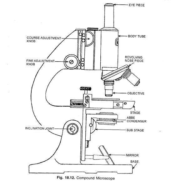
It is another type of optical microscope (the first one being a ‘simple microscope’). Optical parts (a) mechanical parts of a compound microscope. Web a compound microscope is a device used to magnify an object extensively using a combination of lenses. The diaphragm is present at the bottom of the stage. Also, read about the uses of a compound microscope.
Compound Microscope Drawing at GetDrawings Free download
Now visit here to know compound microscope, diagram,. The term compound refers to the usage of more than one lens in the microscope. This stands by resting on the base and supports the stage. Is used in hospitals and forensic labs by scientists, biologists and researchers to study microorganisms. Web microscope parts and functions with labeled diagram and functions how.
Parts Of A Compound Microscope Drawing
Eyepiece (ocular lens) with or without pointer: Use a curved line to enclose a rounded shape beneath the head. The distance between the front of the objective lens and specimen surface when the specimen is focused. Web how to draw compound microscope diagram step by step drawinghow to draw compound microscope#howtodraw #compoundmicroscope #diagrams #abhishekdrawing Web the term “compound” refers to.
Compound Microscope Drawing at Explore collection
Now visit here to know compound microscope, diagram,. The first lens is the objective lens and the second lens is known as the eyepiece lens. Web microscope parts and functions with labeled diagram and functions how does a compound microscope work?. Compound microscopes are built using a compound lens system where the primary magnification is provided by the objective lens,.
Compound Microscope Drawing at Explore collection
The first lens is the objective lens and the second lens is known as the eyepiece lens. Web microscope parts and functions with labeled diagram and functions how does a compound microscope work?. Invented in the late 16th century. It is a vertical projection. Also, read about the uses of a compound microscope.
Compound Microscope Sketch at Explore collection
It is also called a body tube or eyepiece tube. The arm of the microscope is another structural piece. Also, the compound microscope is one of the types of optical microscopes. Notice the bend in the middle of each line. Compound microscopes are built using a compound lens system where the primary magnification is provided by the objective lens, which.
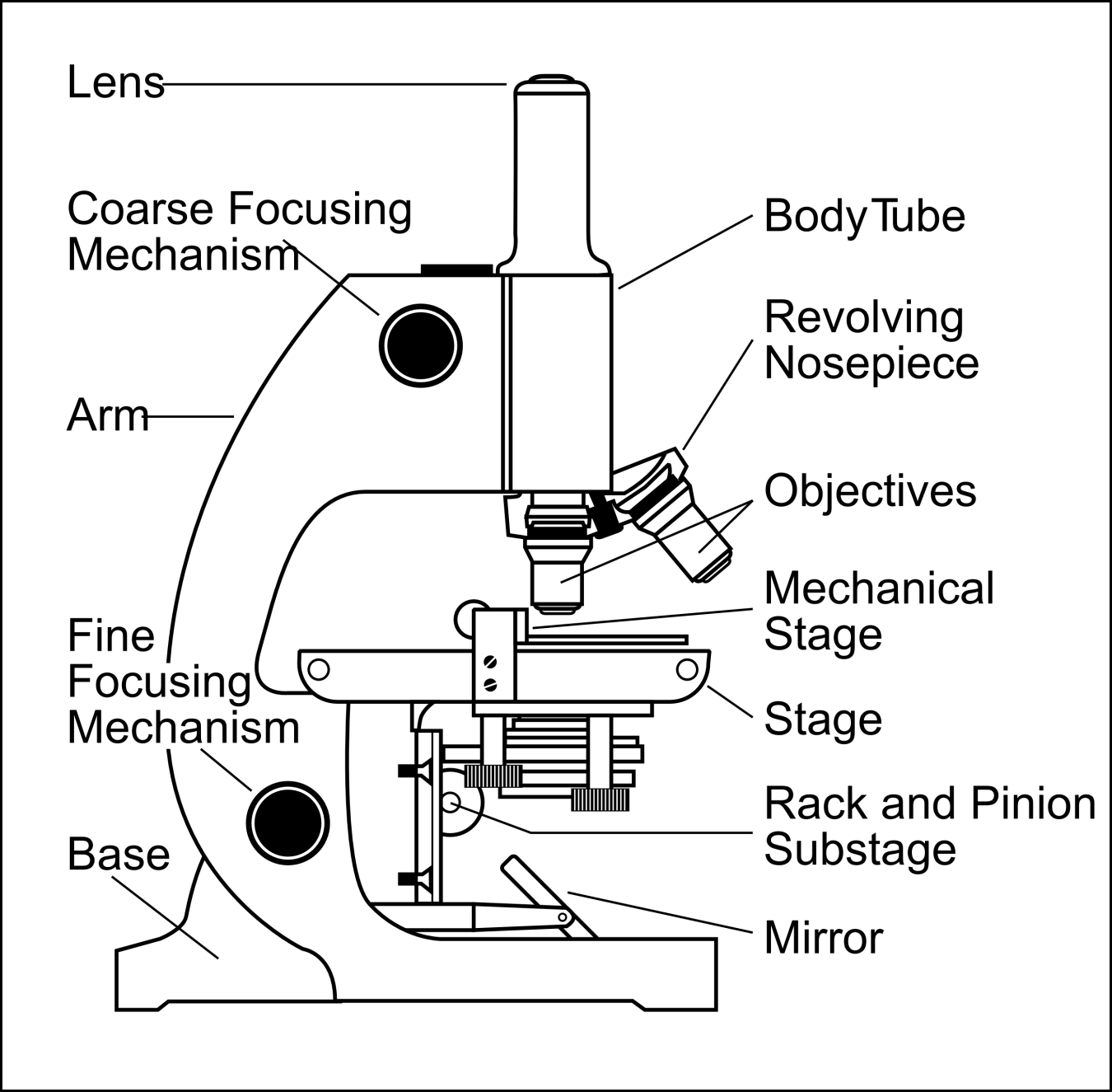
Web Microscope Parts And Functions With Labeled Diagram And Functions How Does A Compound Microscope Work?.
A condenser is an optical tool with which we can focus the light by moving it up and down. Compound microscopes are built using a compound lens system where the primary magnification is provided by the objective lens, which is then compounded (multiplied) by the ocular lens (eyepiece). Is used to view samples that are not visible to the naked eye. Below this, draw another curved line, leaving the shape open on one side.
The Arm Of The Microscope Is Another Structural Piece.
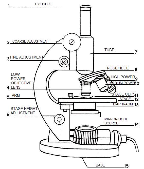
The part that is looked through at the top of the compound microscope. Web how to draw compound microscope diagram step by step drawinghow to draw compound microscope#howtodraw #compoundmicroscope #diagrams #abhishekdrawing Web the parts of the compound microscope can be categorized into: This stands by resting on the base and supports the stage.
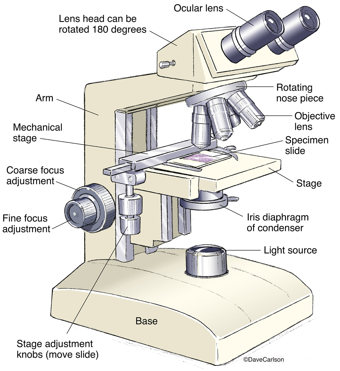
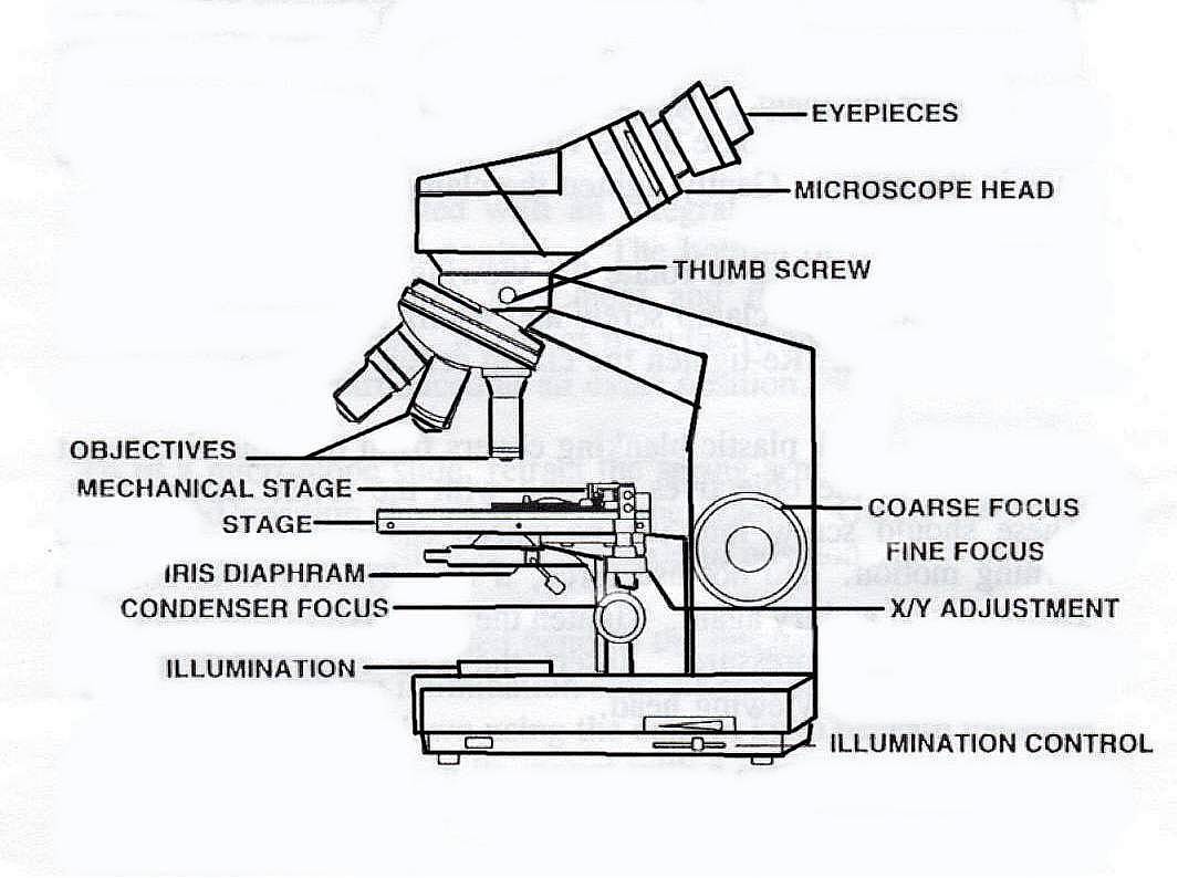
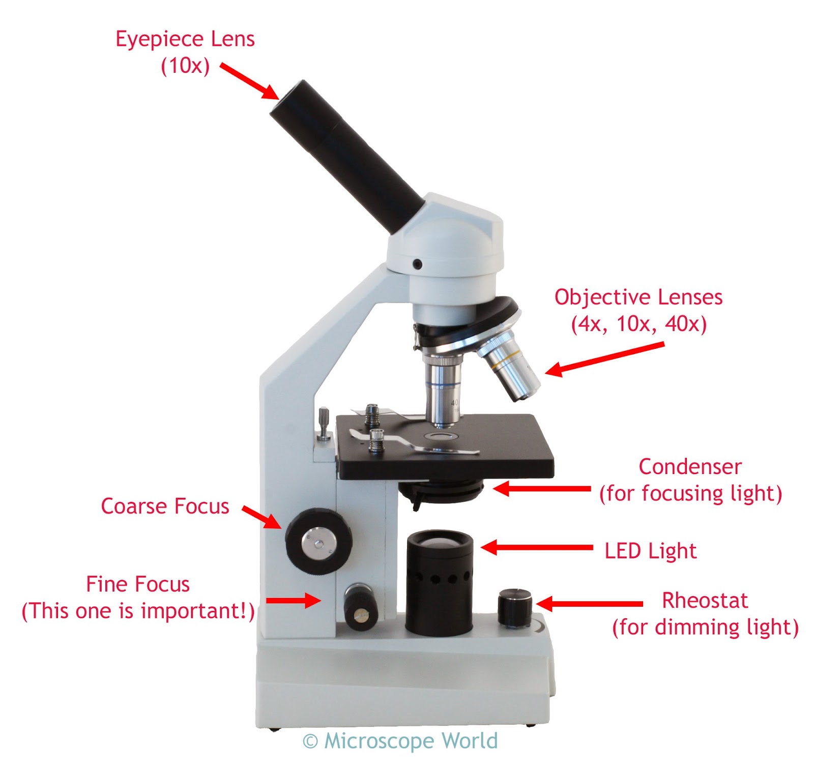
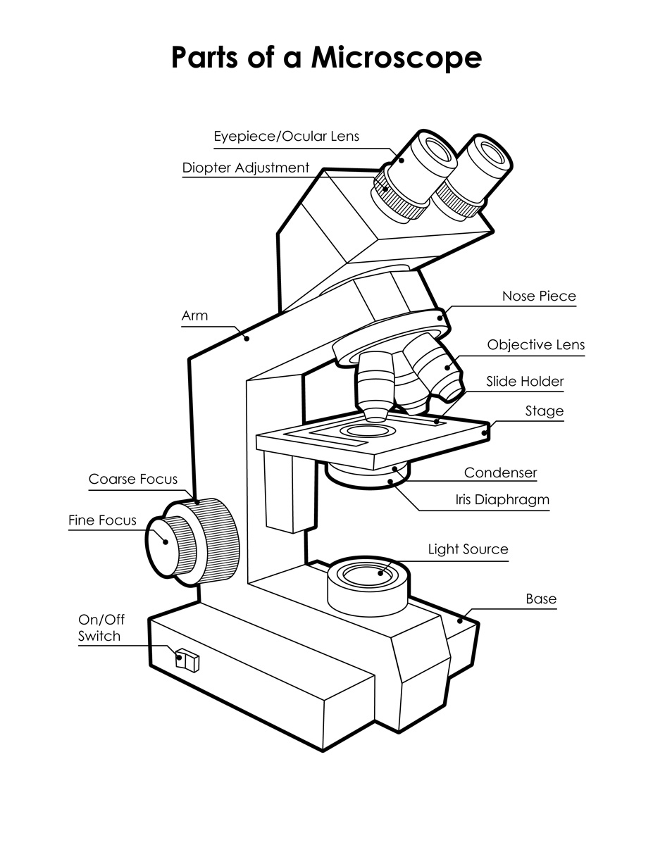
Diagram Of Parts Of A Microscope.
Use a small blending brush and some black paint to create a ground shadow. Web in compound microscopes with two eye pieces there are prisms contained in the body that will also split the beam of light to enable you to view the image through both eye pieces. The orientation of the image you see is flipped in relation to the actual object you’re examining. Web compound microscope definitions for labels.
Now Visit Here To Know Compound Microscope, Diagram,.
The first lens is the objective lens and the second lens is known as the eyepiece lens. The diaphragm is present at the bottom of the stage. Optical parts (a) mechanical parts of a compound microscope. Tutoroot is one of the finest online tutoring platform.