Cranial Drawer Dog
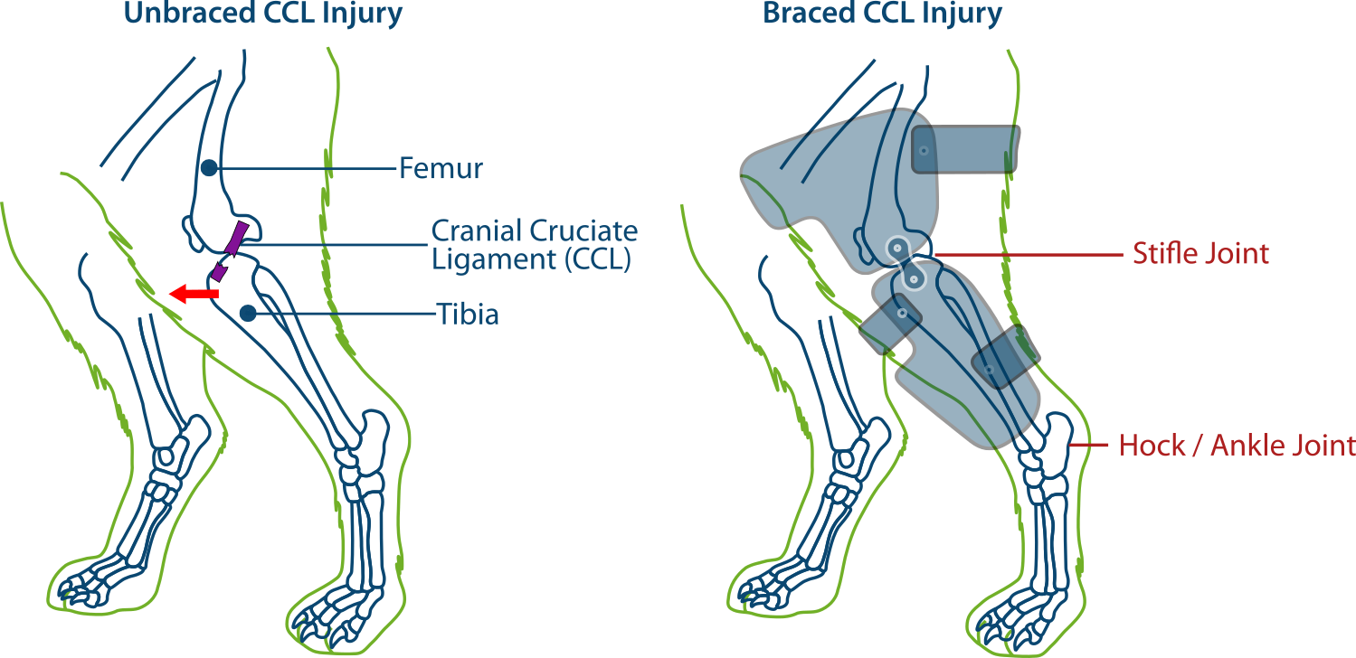
Cranial Drawer Dog - The front part of the tibia is cut and separated from the rest of the tibia. Web cranial cruciate ligament rupture (ccl) is one of the most common orthopedic injuries in dogs. Web both the traditional ecls and the tight rope® procedure are considered extracapsular or external repairs of ccl injury. If this is the case, your veterinarian might recommend giving your dog a sedative to complete the examination. Ccld is the most commonly diagnosed disorder of the canine stifle joint 1 and is the orthopedic disease most commonly treated. Blue = cranial cruciate ligament; When it ruptures, abnormal movement of. Cranial cruciate ligament disease (ccld) is caused by a gradual degeneration of the cranial cruciate ligament (ccl), which results in a partial or complete ligament tear. Web the cranial cruciate ligament (or ccl, see figure 1) is one of the most important stabilizers inside the knee (also called “stifle”) joint, the middle joint in the back leg. Diagnosis of cranial cruciate ligament injuries in dogs by tibial.
Web to test for cranial tibial translation, perform the cranial drawer test (figure 6). It is one of the ligaments that connects the thighbone (femur) to the shinbone (tibia) where they meet at the knee (known as the stifle joint in dogs) and help keep the joint stable. This injury, similar to a torn acl in humans, can come on suddenly or can appear gradually over a longer period. Web the cranial cruciate ligament (or ccl, see figure 1) is one of the most important stabilizers inside the knee (also called “stifle”) joint, the middle joint in the back leg. Web cranial cruciate ligament disease in dogs. Diagnosis of cranial cruciate ligament (ccl) tears is made through a combination of orthopedic examination findings (eg, positive cranial drawer, cranial tibial translation) and radiographic changes (eg, effusion, osteoarthritic change). Web testing for cranial drawer or tibial thrust is not painful; In dogs, the most common knee injury is a rupture or tear of the cranial cruciate ligament. Two cruciate ligaments, the cranial (anterior) and the posterior cruciate ligaments, are found in the knee joint of dogs and cats (and most other domestic animals). In humans the ccl is called the anterior cruciate ligament (acl).
Web both the traditional ecls and the tight rope® procedure are considered extracapsular or external repairs of ccl injury. Web cranial cruciate ligament rupture in dogs. Web cranial cruciate ligament disease in dogs. Web a positive tibial compression test and cranial drawer test confirm cclr. When it ruptures, abnormal movement of. Veterinary school instruction has traditionally emphasized teaching subtle and difficult manipulative physical examination procedures, such as cranial drawer sign and cranial tibial thrust, to definitively diagnose crclr.cranial tibial thrust is the stifle vector. It is one of the ligaments that connects the thighbone (femur) to the shinbone (tibia) where they meet at the knee (known as the stifle joint in dogs) and help keep the joint stable. Diagnosisdiagnosis is made by elicitation of positive cranial drawer or cranial tibial thrust or by palpation of medial buttress.1 medial buttress occurs rapidly after ligament rupture, and is ample indication for surgical joint exploration even in the absence of laxity. Cranial cruciate ligament disease (ccld) is caused by a gradual degeneration of the cranial cruciate ligament (ccl), which results in a partial or complete ligament tear. Web the cranial drawer test and tibial compression tests are important for assessing palpable instability.

Dog with Cranial Drawer YouTube
Web the rupture of the cranial cruciate ligament allows the tibia to slide forward. Diagnosisdiagnosis is made by elicitation of positive cranial drawer or cranial tibial thrust or by palpation of medial buttress.1 medial buttress occurs rapidly after ligament rupture, and is ample indication for surgical joint exploration even in the absence of laxity. During this exam your veterinary physician.
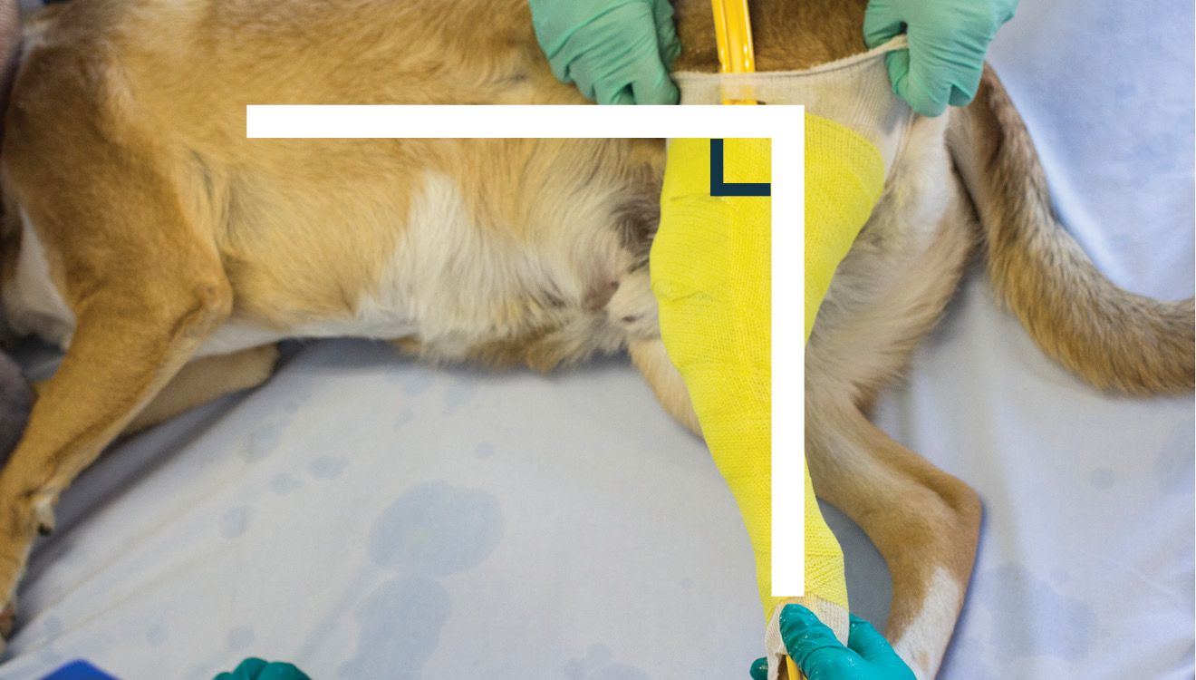
4 Tips To The Perfect Dog Brace Cast Hero Blog
The front part of the tibia is cut and separated from the rest of the tibia. The examiner stands behind the dog and places a thumb on the caudal aspect of the femoral. (1) prevent cranial displacement of the tibia in relation to the femur (cranial drawer sign) (2) prevent hyperextension of the knee, and (3) prevent internal rotation of.
Torn ACL in Dogs How Braces Help
Web the cranial cruciate ligament (ccl) in dogs is the equivalent of the anterior cruciate ligament (acl) in humans. The examiner stands behind the dog and places a thumb on the caudal aspect of the femoral. Veterinary school instruction has traditionally emphasized teaching subtle and difficult manipulative physical examination procedures, such as cranial drawer sign and cranial tibial thrust, to.

Pathology, Diagnosis, and Treatment Goals of Cranial Cruciate Ligament
The cranial cruciate ligament (ccl) is responsible for stabilizing the tibia from abnormally thrusting forward and away from the femur. Ccld is the most commonly diagnosed disorder of the canine stifle joint 1 and is the orthopedic disease most commonly treated. Diagnosisdiagnosis is made by elicitation of positive cranial drawer or cranial tibial thrust or by palpation of medial buttress.1.
Ruptured cruciate ligaments in dogs which is the best surgery? Vet
In dogs with a ruptured cranial cruciate ligament, the tibia will display forward motion upon flexion of the ankle joint. Humans have a similar anatomical structure to the dog's knee, but the ligaments are called the anterior and posterior cruciate ligaments. Therefore, a positive cranial drawer test is indicative of cruciate ligament damage. (1) prevent cranial displacement of the tibia.

Cruciate Disease The Cranial Drawer Test YouTube
Web the cranial cruciate ligament (ccl) in dogs is the equivalent of the anterior cruciate ligament (acl) in humans. Ccld is the most commonly diagnosed disorder of the canine stifle joint 1 and is the orthopedic disease most commonly treated. Web to test for cranial tibial translation, perform the cranial drawer test (figure 6). Web cranial cruciate ligament rupture (ccl).

Cranial Cruciate Ligament Dogs
Humans have a similar anatomical structure to the dog's knee, but the ligaments are called the anterior and posterior cruciate ligaments. The examiner stands behind the dog and places a thumb on the caudal aspect of the femoral. However, some dogs might be too tense or nervous to allow a thorough exam. In humans the crcl is called the anterior.

Positive cranial drawer sign in a dog with a cranial (anterior
The cruciate ligaments are tough fibrous bands that connect the distal femur (thigh bone) to the proximal tibia (shin bone). Diagnosisdiagnosis is made by elicitation of positive cranial drawer or cranial tibial thrust or by palpation of medial buttress.1 medial buttress occurs rapidly after ligament rupture, and is ample indication for surgical joint exploration even in the absence of laxity..
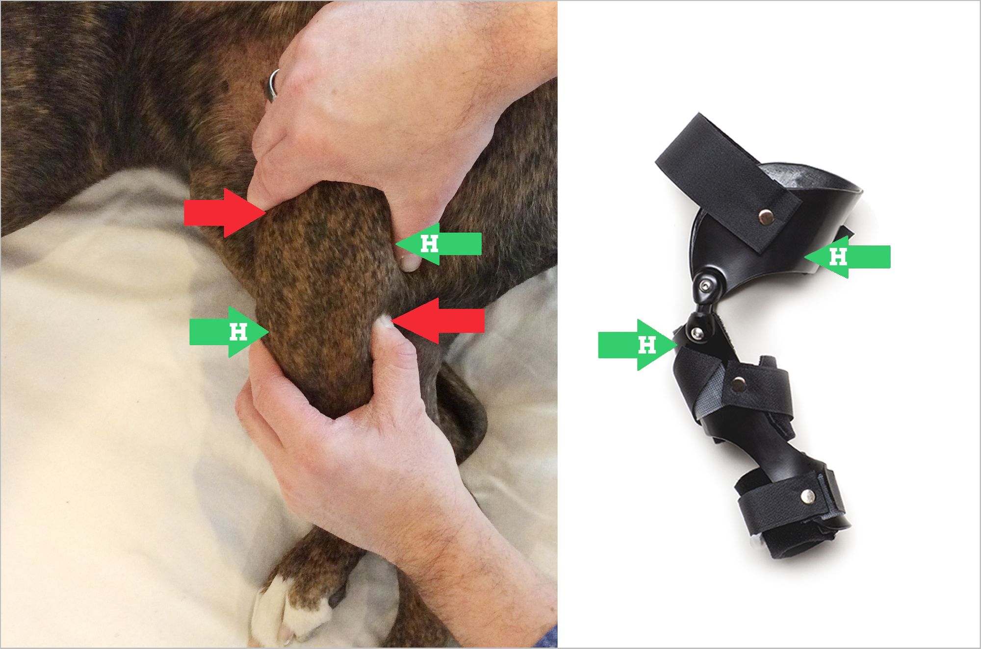
Dog Stifle CCL/ACL Injury Support Brace — PawOpedic
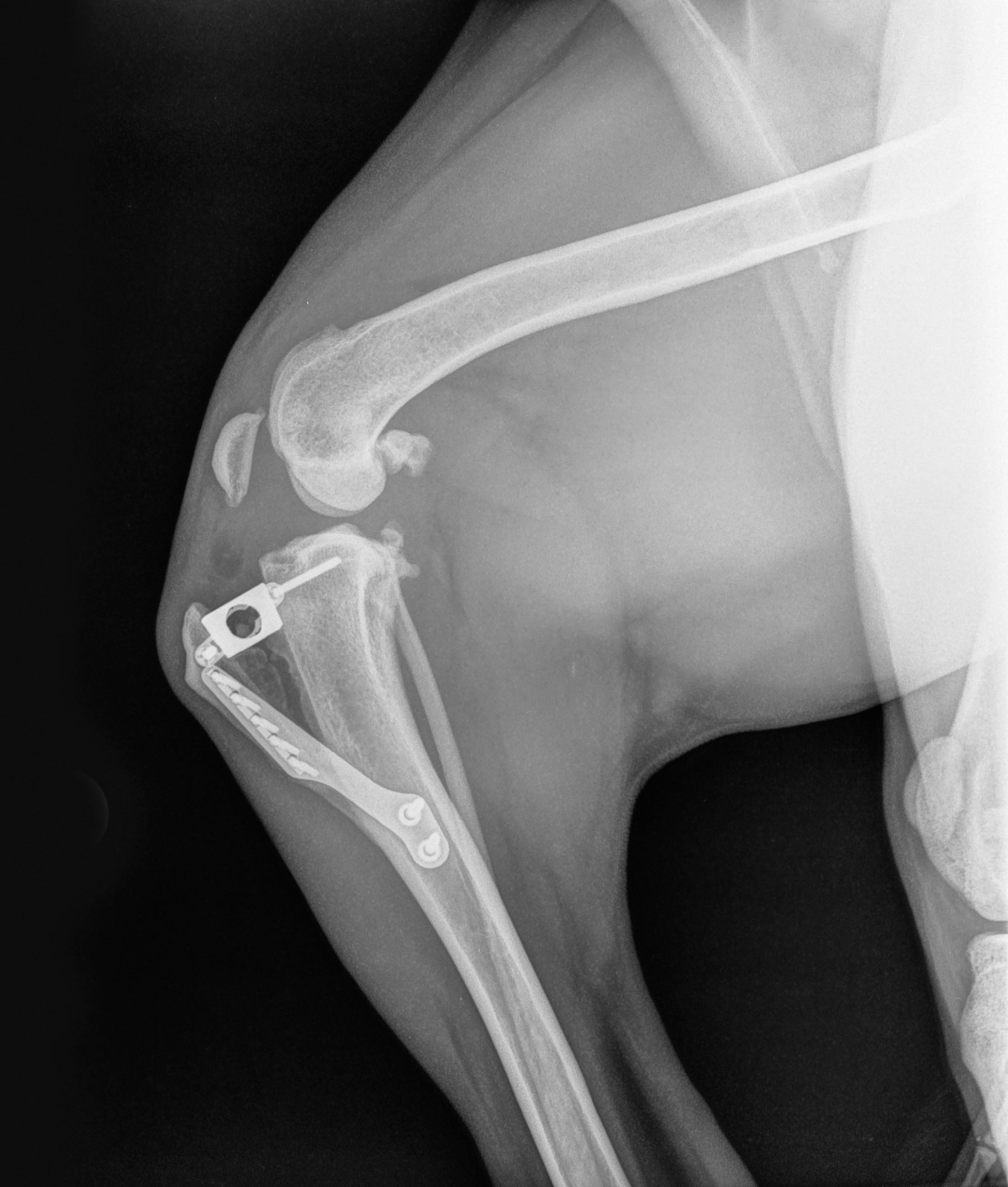
In humans the crcl is called the anterior cruciate ligament (acl). For this reason, i prefer to perform a tibial compression test for evaluation of cranial tibial thrust instability at the conclusion of my. The cranial cruciate ligament (ccl) is responsible for stabilizing the tibia from abnormally thrusting forward and away from the femur. A special orthopedic spacer is screwed.

Cranial Suture Closure in Domestic Dog Breeds and Its Relationships to
Web another test, similar to the eliciting the drawer sign, is the tibial compression test. Web to test for cranial tibial translation, perform the cranial drawer test (figure 6). If this is the case, your veterinarian might recommend giving your dog a sedative to complete the examination. A special orthopedic spacer is screwed into the space between the two sections.
Web The Canine Cranial Cruciate Ligament Is One Of Four Ligaments That Join The Tibia And Femur Together To Create A Stable Dog Knee Joint (Stifle Joint).
This motion is just like pulling a drawer open. Humans have a similar anatomical structure to the dog's knee, but the ligaments are called the anterior and posterior cruciate ligaments. In dogs with a ruptured cranial cruciate ligament, the tibia will display forward motion upon flexion of the ankle joint. Diagnosisdiagnosis is made by elicitation of positive cranial drawer or cranial tibial thrust or by palpation of medial buttress.1 medial buttress occurs rapidly after ligament rupture, and is ample indication for surgical joint exploration even in the absence of laxity.
During This Exam Your Veterinary Physician Will Stabilize The Dog’s Femur With One Hand, While Flexing The Ankle With The Other.
Ccld is the most commonly diagnosed disorder of the canine stifle joint 1 and is the orthopedic disease most commonly treated. Unlike human athletes, rupture of the ccl in dogs is rarely the result of a traumatic injury. Van ryssen b, van bree h. When it ruptures, abnormal movement of.
Some Dogs Are More Relaxed In The Standing Position Than When Restrained In Lateral Recumbency.
However, some dogs might be too tense or nervous to allow a thorough exam. Web testing for cranial drawer or tibial thrust is not painful; Web the cranial cruciate ligament (ccl) in dogs is the equivalent of the anterior cruciate ligament (acl) in humans. Web the cranial cruciate ligament (crcl, see figure 1.) is one of the most important stabilizers inside the canine knee (stifle) joint, the middle joint in the back leg.
It Is One Of The Ligaments That Connects The Thighbone (Femur) To The Shinbone (Tibia) Where They Meet At The Knee (Known As The Stifle Joint In Dogs) And Help Keep The Joint Stable.
Web in dogs and cats, the ligaments are called the cranial and caudal cruciate ligaments. The two primary risks of extracapsular surgical repairs are infection and failure. The examiner stands behind the dog and places a thumb on the caudal aspect of the femoral. Diagnosis of cranial cruciate ligament (ccl) tears is made through a combination of orthopedic examination findings (eg, positive cranial drawer, cranial tibial translation) and radiographic changes (eg, effusion, osteoarthritic change).