Dermis Drawing
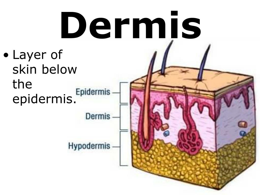
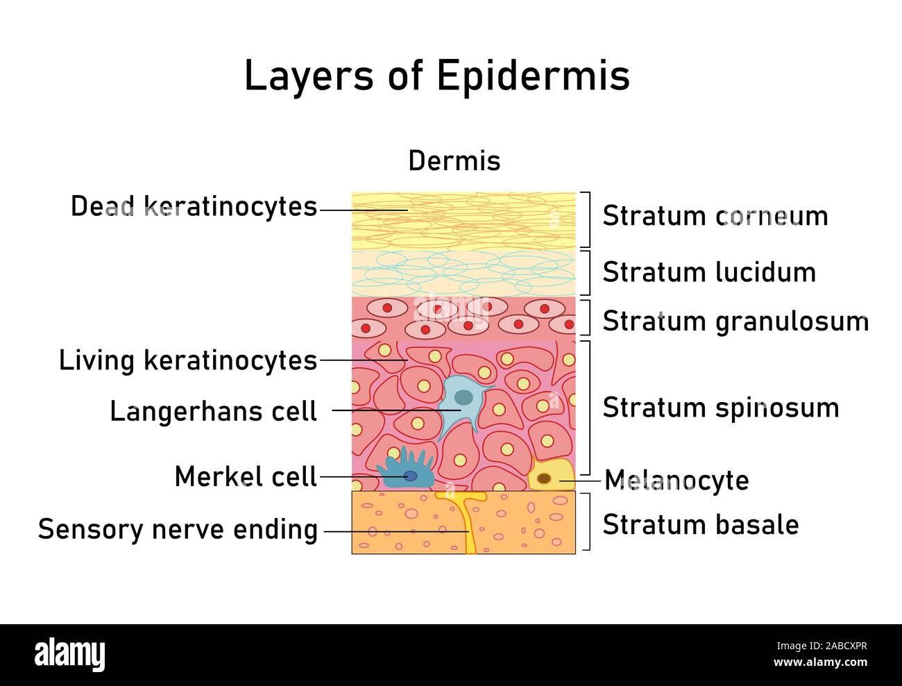
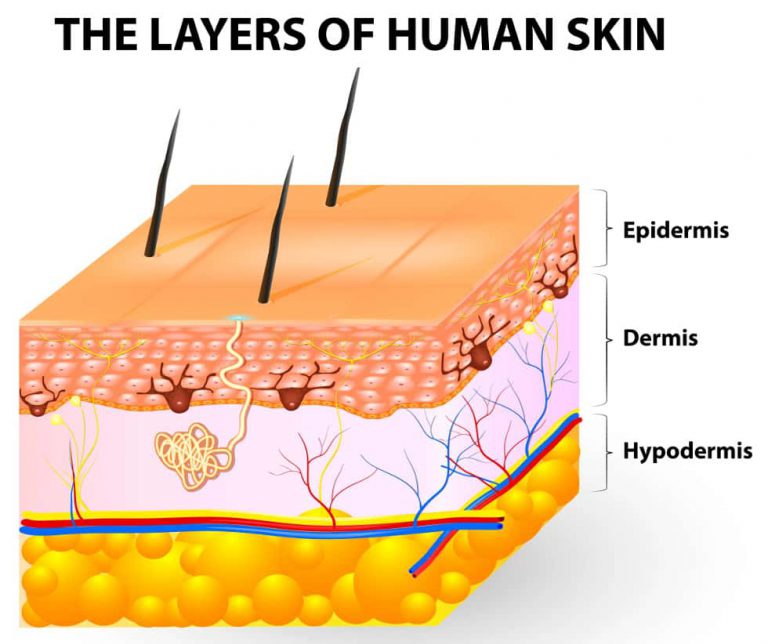
Dermis Drawing - Web the dermis has two parts: The stratum corneum is the top layer of the epidermis. Also shown are the hair shafts, hair follicles, oil glands, lymph vessels, nerves, fatty. The university of waikato te whare wānanga o waikato published 1 february 2011 size: This involves increased keratin production and migration toward the external surface, a process termed cornification. Web the dermis or corium is a layer of skin between the epidermis (with which it makes up the cutis) and subcutaneous tissues, that primarily consists of dense irregular connective tissue and cushions the body from stress and strain. (dermis and hypodermis) google classroom. Find out more about its structure and function at kenhub! Drawing shows the epidermis (including the squamous cell and basal cell layers), dermis, and subcutaneous tissue. It is made up of the following five layers.
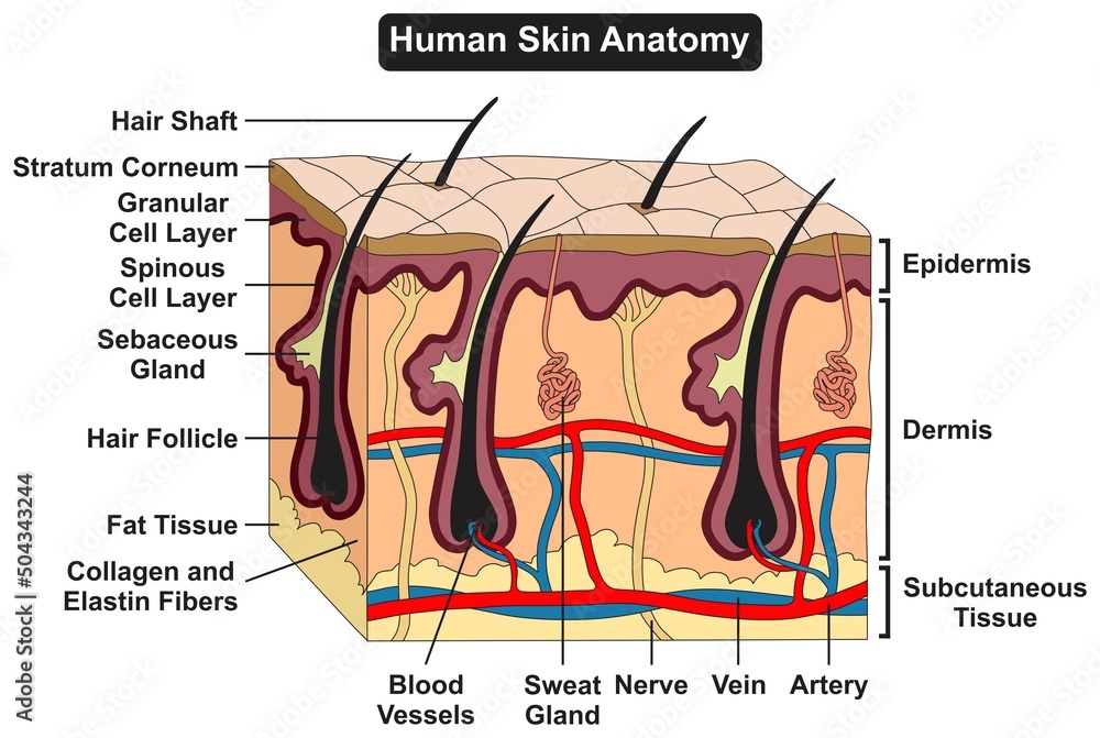
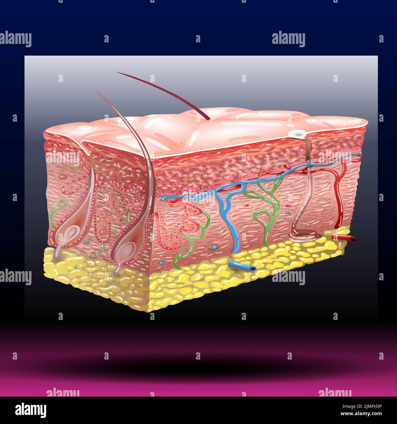
Web the dermis has two parts: Web diagram of human skin structure. How big is the dermis? A thin, upper layer known as the papillary dermis, and a thick, lower layer known as the reticular dermis. Find out more about its structure and function at kenhub! This article will describe the anatomy and histology of the skin. Understand the different types of tissues, their functions, and how they contribute to our sensory experiences. Keep unwanted substances out of your body. Nerve plexus around hair follicle. Differentiate among the regions of the dermis and the hypodermis.
It has only two layers: It is made of dead, flattened cells called keratinocytes that are shed approximately every two. Ruffini ending (terminal) sebaceous gland. New 3d rotate and zoom. Find out more about its structure and function at kenhub! The primary function of the dermis is to cushion the body from stress and strain, and to also provide: What lies beneath the epidermis? Web atopic dermatitis (eczema) plaque psoriasis. This involves increased keratin production and migration toward the external surface, a process termed cornification. On the back, the palms of hands and the soles of feet, it measures 3 millimeters thick
PPT 7th Grade Unit 5 The Structure and Function of Body Systems
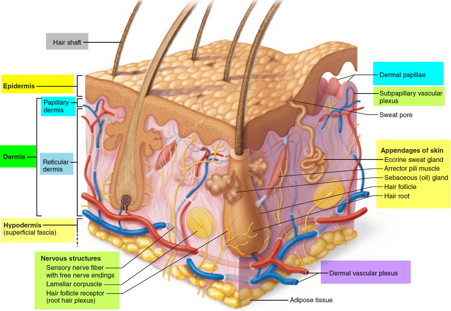
Web the dermis is divided into a papillary region and a reticular region. Elasticity to the skin, a sense of touch, and heat. Skin [38:23] general histology of the skin. For example, the dermis on the eyelids is 0.6 millimeters thick; Nevus (birthmark, mole, or “port.
Dermis Layers, Papillary Layer, Function Epidermis
Its thickness varies depending on the location of the skin. Web atopic dermatitis (eczema) plaque psoriasis. It contains blood and lymph vessels, nerves, and other structures, such as hair follicles and sweat glands. Melanocytes that produce melanin (influences skin color), keratinocytes that produce keratin, merkel’s cells that function in touch, and langerhans’ cells that function in. Ruffini ending (terminal) sebaceous.
Vector illustration with structure of dermis for medical and
The dermis is divided into a papillary region and a reticular region. Web (dermis and hypodermis) (video) | khan academy. Understand the different types of tissues, their functions, and how they contribute to our sensory experiences. Drawing shows layers of the epidermis, dermis, and subcutaneous tissue including hair shafts and follicles, oil glands, lymph vessels, nerves, fatty tissue, veins, arteries,.

Dermis layers Dermis, Layers, Dermal fillers
Also shown are the hair shafts, hair follicles, oil glands, lymph vessels, nerves, fatty. This involves increased keratin production and migration toward the external surface, a process termed cornification. Your dermis varies in thickness across your body. The dermis contains hair roots, sebaceous glands, sweat glands, nerves, and blood vessels. For example, the dermis on the eyelids is 0.6 millimeters.
Human skin anatomy structure and parts infographic diagram epidermis
The dermis contains hair roots, sebaceous glands, sweat glands, nerves, and blood vessels. Web diagram of human skin structure. It is made of dead, flattened cells called keratinocytes that are shed approximately every two. What is the dermis’s structure? Web the dermis is the layer of skin found deep to the epidermis and superficial to the hypodermis.
TheLayersofhumanskinepidermisdermishypodermis swimfolk
Differentiate among the regions of the dermis and the hypodermis. This article will describe the anatomy and histology of the skin. It contains blood and lymph vessels, nerves, and other structures, such as hair follicles and sweat glands. View this animation to learn more about layers of the skin. Your dermis varies in thickness across your body.

Anatomy of human skin. The most superficial layer of the skin is the
There are four types of cells that make up the epidermis: It is made of dead, flattened cells called keratinocytes that are shed approximately every two. Web your dermis is the middle layer of your skin, located between your epidermis (top layer) and hypodermis (bottom layer) in your skin. Nevus (birthmark, mole, or “port. For example, the dermis on the.
Skin anatomy. Human normal skin dermis epidermis adipose layers recent
Web (dermis and hypodermis) (video) | khan academy. Skin [38:23] general histology of the skin. Web the dermis connects the epidermis to the hypodermis, and provides strength and elasticity due to the presence of collagen and elastin fibers. Web the dermis has two parts: Drawing shows layers of the epidermis, dermis, and subcutaneous tissue including hair shafts and follicles, oil.

Structure of the epidermis medical vector illustration, dermis anatomy
B&w, medical illustration (jpeg format) source: Web (dermis and hypodermis) (video) | khan academy. What is the dermis’s structure? It is made up of the following five layers. Web this video explains what the dermis is and explains the components as well as the function of the dermissupport us!:

The structure of the skin is composed of two layers (1) the epidermis
Skin [38:23] general histology of the skin. The skin consists of two main layers and a closely associated layer. The stratum corneum is the top layer of the epidermis. Your dermis varies in thickness across your body. On the back, the palms of hands and the soles of feet, it measures 3 millimeters thick
New 3D Rotate And Zoom.
Web your dermis is the middle layer of your skin, located between your epidermis (top layer) and hypodermis (bottom layer) in your skin. ‘skin diagram || how to draw and label the parts of skin’ is demonstrated in this video tutorial step by step. What lies beneath the epidermis? Web the dermis consists of a papillary and a reticular layer that serve to protect and cushion the body from stress and strain.
Skin [38:23] General Histology Of The Skin.
This involves increased keratin production and migration toward the external surface, a process termed cornification. Web the dermis has two parts: Web beneath the dermis lies the hypodermis, which is composed mainly of loose connective and fatty tissues. Web the dermis or corium is a layer of skin between the epidermis (with which it makes up the cutis) and subcutaneous tissues, that primarily consists of dense irregular connective tissue and cushions the body from stress and strain.
Uruj Zehra, Mbbs, Mphil, Phd.
Your dermis varies in thickness across your body. Its thickness varies depending on the location of the skin. Keep unwanted substances out of your body. The dermis contains hair roots, sebaceous glands, sweat glands, nerves, and blood vessels.
Web Atopic Dermatitis (Eczema) Plaque Psoriasis.
B&w, medical illustration (jpeg format) source: Explore the complex layers of skin, from the epidermis to the hypodermis. The epidermis is the most superficial layer of the skin, and is largely formed by layers of keratinocytes undergoing terminal maturation. There are four types of cells that make up the epidermis: