Draw A Human Epithelial Cell And An Elodea Cell
Draw A Human Epithelial Cell And An Elodea Cell - As you can see in the image, the shapes of the cells vary to some degree, so taking an average of three cells’ dimensions, or even the results from the entire class, gives a more accurate determination of. Wet mount of human epithelial cell (cheek cell) exercise 6: Review the major organelles of eukaryotes; Wet mount of an elodea leaf cell. This human cheek cell is a good example of a typical animal cell. List how the animal cells differ from the plant cells. Discover how chloroplasts while undergoing photosynthesis; Cells with smaller sizes and relatively smaller sizes compared to their neighbors exhibit an increased likelihood of undergoing apoptosis. Web the onion and elodea cells are interconnected like a brick formation, whereas cheek cells are just overlapping and kind of near to each other. It has a prominent nucleus and a flexible cell membrane which.
You probably will not see the cells at this. Web in this lab you will look at two types of cells, a human cheek cell and an elodea cell and see how they are similar and how they are different. Review the major organelles of eukaryotes; How many nucleoli are present in each nucleus? Discover how chloroplasts while undergoing photosynthesis; Elodea is a water plant that grows abundantly in ponds around spokane. As you can see in the image, the shapes of the cells vary to some degree, so taking an average of three cells’ dimensions, or even the results from the entire class, gives a more accurate determination of. Place the elodea leaf into the drop of water on your slide. While observing the leaf under the microscope, wick a solution of 6% nacl (sodium chloride) across the slide. Web paper towels or tissues.
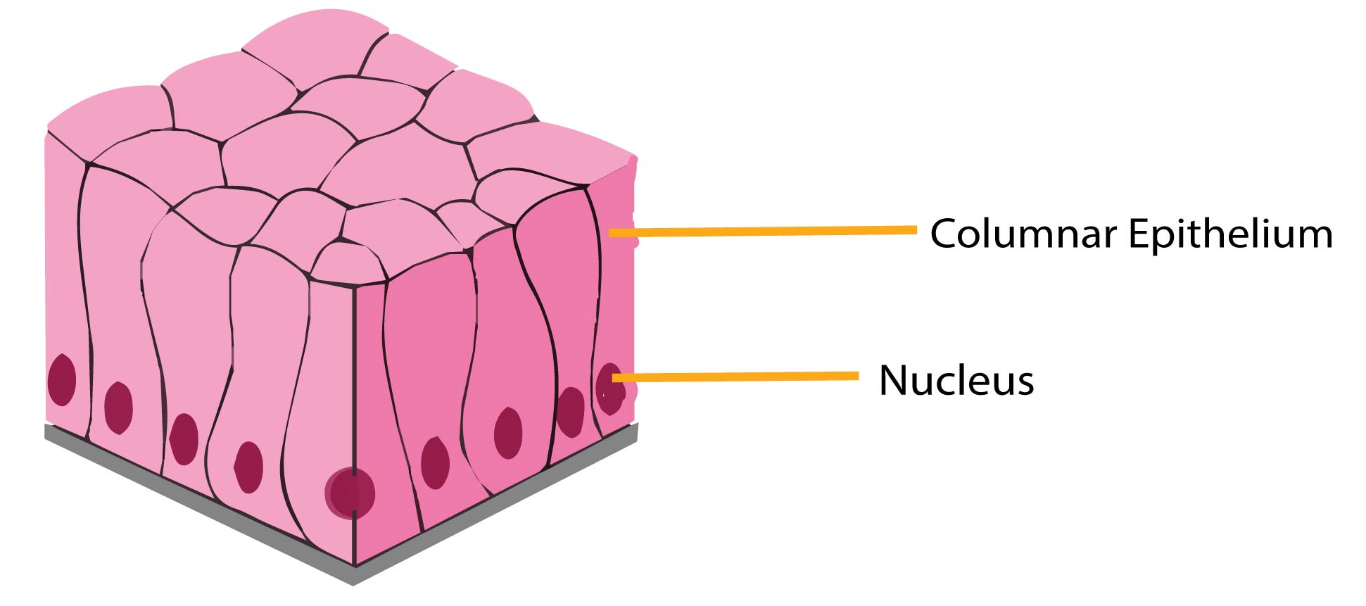
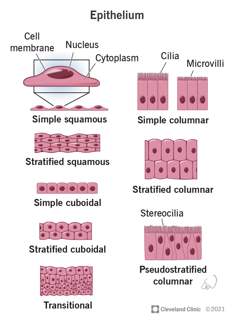
Observe the structures of elodea, alium and human cells. Prepare a wet mount of one leaf from the water plant elodea using the water in which it is kept. Distinguish between simple epithelia and stratified epithelia, as well as between squamous, cuboidal, and. Web the human cheek is lined with epithelial cells. Draw three representative cells, each about 2 cm in diameter. A cuboidal epithelial cell looks close to a square. Structures found on some epithelial cells are an adaptation to specific functions. Web human epithelial cells and elodea cells differ in a number of ways. First of all, elodea cells are only found in plants while epithelial cells are. You probably will not see the cells at this.

Diagram Of Elodea Cell
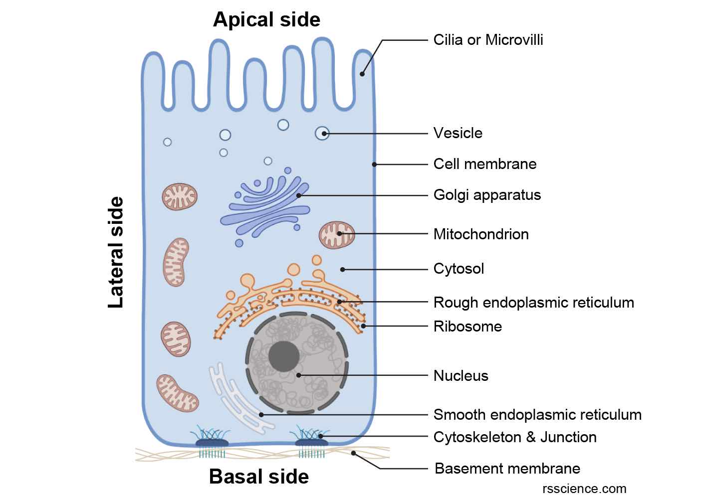
Structures found on some epithelial cells are an adaptation to specific functions. Identify easily identifiable organelles within human cells; Place a coverslip onto the slide. Explain the structure and function of epithelial tissue. Human cells tend to be a little.

How Do Human Epithelial Cells And Elodea Cells Differ HOWDOZD
Web a “typical” elodea cell is approximately 0.05 millimeters long (50 micrometers long) and 0.025 millimeters wide (25 micrometers wide). First of all, elodea cells are only found in plants while epithelial cells are. Web in the lab the cell of the biology lab primer, you will: Observe the structures of elodea, alium and human cells. As you can see.

Diagram Of Elodea Cell
Label the structures in one cell: Cytoplasm, nucleus, cell wall, plasma membrane, vacuole, and chloroplasts. Distinguish between tight junctions, anchoring junctions, and gap junctions. Despite more than half a century of research, mscs continue to be among the most extensively studied cell types in. How does the shape of the elodea cells differ from that of the cheek cells?
Describe various types of epithelial tissues with the help of labeled
Place the elodea leaf into the drop of water on your slide. Human cells tend to be a little. Draw three representative cells, each about 2 cm in diameter. As you can see in the image, the shapes of the cells vary to some degree, so taking an average of three cells’ dimensions, or even the results from the entire.
[Solved] 3 Draw a human epithelial cell and an Elodea cell. Label the
While observing the leaf under the microscope, wick a solution of 6% nacl (sodium chloride) across the slide. Prepare cheek cell and plant cell slide. What visible structures do the elodea and the onion skin cells share? How does the shape of the elodea cells differ from that of the cheek cells? Wet mount of an elodea leaf cell.
Epithelium What It Is, Function & Types
As you can see in the image, the shapes of the cells vary to some degree, so taking an average of three cells’ dimensions, or even the results from the entire class, gives a more accurate determination of. Wet mount of human epithelial cell (cheek cell) exercise 6: Place a coverslip onto the slide. A columnar epithelial cell looks like.
Epithelium Definition, Characteristics, Cell Structures, Types, and
Water will flow out of the elodea cells by osmosis, shrinking the cell membrane away from the stiff cell wall (plasmolysis). As you can see in the image, the shapes of the cells vary to some degree, so taking an average of three cells’ dimensions, or even the results from the entire class, gives a more accurate determination of. Label.
[Solved] 3 Draw a human epithelial cell and an Elodea cell. Label the
Web find the cell membrane, nucleus, nuclear envelope, and cytoplasm. Illustrations of how to prepare a wet mount slide (mader 2001). What purpose do epithelial cells serve? Figure 2 human cheek cells. Explain the structure and function of epithelial tissue.
Epithelial Tissues And Their Functions Anatomy
Put a drop of water onto the microscope slide. How does the shape of the elodea cells differ from that of the cheek cells? Identify easily identifiable organelles within human cells; Structures found on some epithelial cells are an adaptation to specific functions. A cuboidal epithelial cell looks close to a square.
[Solved] 3 Draw a human epithelial cell and an Elodea cell. Label the
Despite more than half a century of research, mscs continue to be among the most extensively studied cell types in. Review the major organelles of eukaryotes; This cell was alive and at 1000x magnification when it was photographed. Observation of plant cells (elodea cells) exercise 7: Prepare cheek cell and plant cell slide.
Web The Human Cheek Is Lined With Epithelial Cells.
Web human epithelial cells and elodea cells differ in a number of ways. Cells with smaller sizes and relatively smaller sizes compared to their neighbors exhibit an increased likelihood of undergoing apoptosis. This human cheek cell is a good example of a typical animal cell. Web human epithelial cells differ in shape and size from those of onion and elodea cells, which are types of plant cells.
Observe The Cells Under Normal Conditions, And Make A Sketch Of What You See.
They will be used today for you to observe a eukaryotic animal cells and its nucleus. You probably will not see the cells at this. Put a drop of water onto the microscope slide. Cytoplasm, nucleus, cell wall, plasma membrane, vacuole, and chloroplasts.
A Micrograph Of A Cell Nucleus.
What purpose do epithelial cells serve? 8match the following functions with their corresponding structure: As you can see in the image, the shapes of the cells vary to some degree, so taking an average of three cells’ dimensions, or even the results from the entire class, gives a more accurate determination of. Calibrate a microscope and determine the size of cells.
How Does The Shape Of The Elodea Cells Differ From That Of The Cheek Cells?
Draw a cell from the azolla in the space below. Prepare cheek cell and plant cell slide. This cell was alive and at 1000x magnification when it was photographed. While observing the leaf under the microscope, wick a solution of 6% nacl (sodium chloride) across the slide.