Draw Bacteria
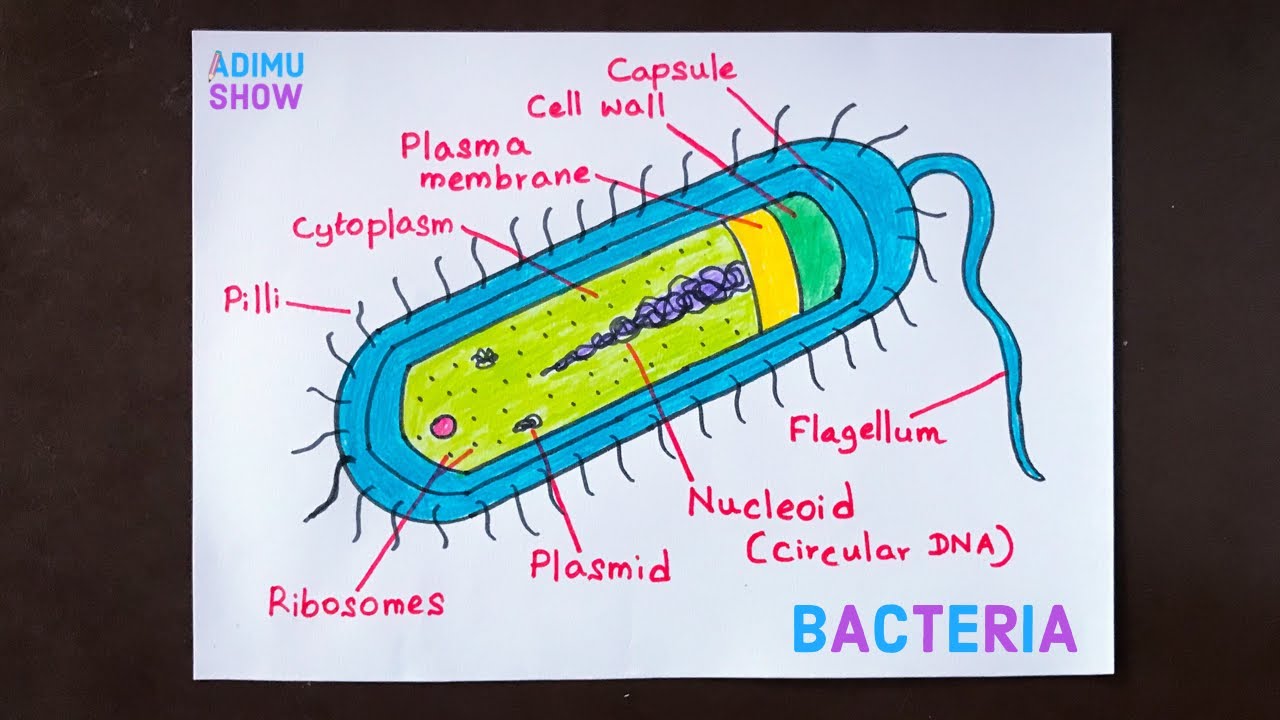
Draw Bacteria - They are also very versatile organisms, surviving in extremely inhospitable conditions. Web bacteria such as clostridium difficile can become problematic and is now a serious type of nosocomial infection. The take home message is that [antibiotics increase selective pressure on bacteria which speed their evolution and create more unique and potentially pathogenic strains, for which our immune system cannot keep pace.] It includes the cell wall of bacteria and the plasma membrane beneath it. Now let's moveon to diagram. The size of these cells range between 1 micrometre to 3 micrometres, so they are barely visible under the light microscope. Web the cytoplasm of prokaryotic cells lacks in well defined cell organelles such as endoplasmic reticulum, golgi apparatus, mitochondria,centrioles, nucleoli, cytoskeleton. Web in this video, we show you how to draw and label a basic bacterial cell. So in this diagram we try t. The capsule offers protection from a variety of different threats to the cell, such as desiccation, hydrophobic toxic materials (i.e.
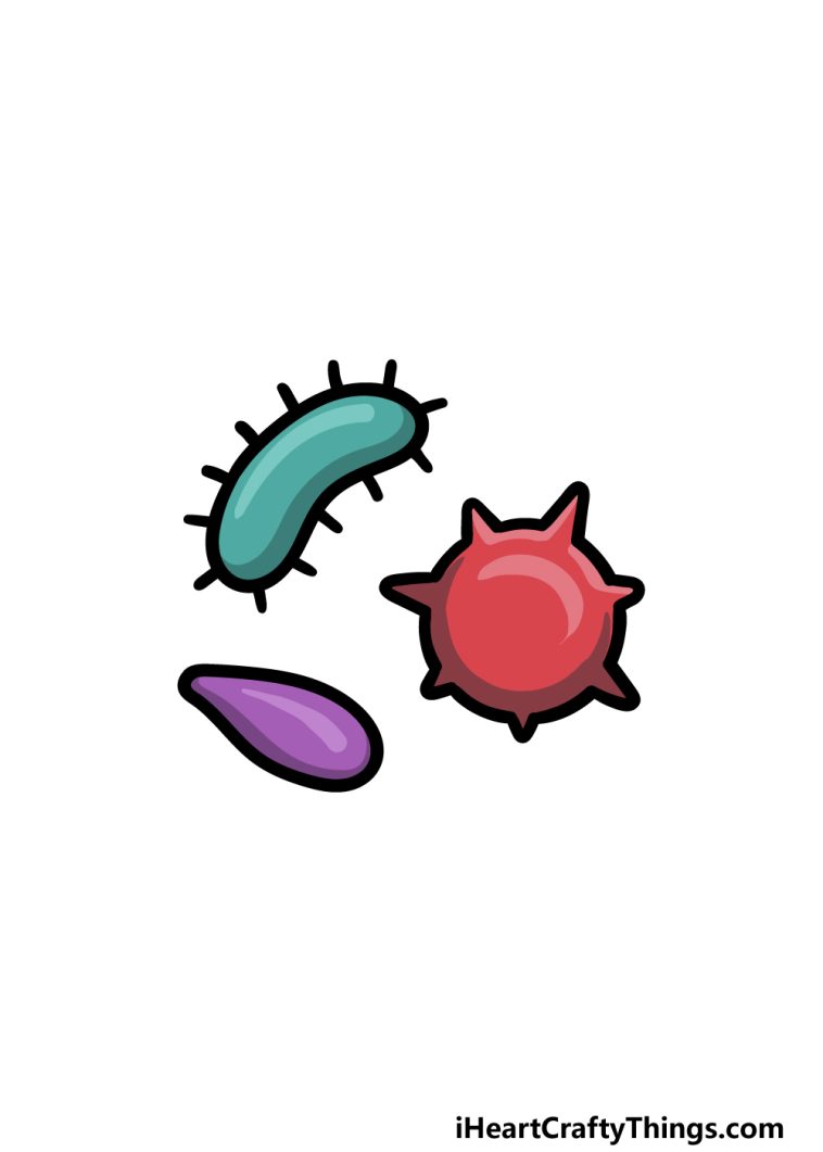
Free & simple drawing tutorials for kindergarten. The bacteria uses its flagellum to swim or move around. The limit of resolution with the unaided eye is about 200 microns, and as many bacteria are smaller than this size, they are not visible with naked eyes. Web the three main shapes of bacteria are coccus, spiral, and bacillus. Now let's moveon to diagram. So in this diagram we try t. Web bacteria such as clostridium difficile can become problematic and is now a serious type of nosocomial infection. Web how to draw bacteria.easy outline diagrambuy the material i used 1. When you have a toothache or tooth infection, keeping your head elevated can help prevent any blood and bacteria from pooling in your mouth, dr. On average, the size of bacteria ranges from 0.5 to 5 µm.
Larger bacterial cells may be visible. A bacterial capsule is a polysaccharide layer that completely envelopes the cell. On average, the size of bacteria ranges from 0.5 to 5 µm. Web the structure of bacteria is known for its simple body design. They are also very versatile organisms, surviving in extremely inhospitable conditions. Web learn step by step drawing tutorial. Let's draw a bacteria in illustrator for your graphical abstract.🎨 drawbiomed is a channel for scientists to learn professional scientific. It includes the cell wall of bacteria and the plasma membrane beneath it. So in this diagram we try t. The bacteria uses its flagellum to swim or move around.

Bacteria Drawing at GetDrawings Free download
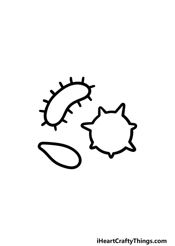
The outer envelope acts as a structural and physiological. We will be keeping things nice and simple for this first part of your bacteria drawing. A typical bacterial cell is structurally very similar to a plant cell. When you have a toothache or tooth infection, keeping your head elevated can help prevent any blood and bacteria from pooling in your.

How To Draw Different Shapes Of Bacteria YouTube
The copy numbers of these proteins range from a few to. This diagram depicts the numerous shapes of bacteria. Web bacteria is a unicellular prokaryotic organism. Such organisms are called extremophiles. The 70s ribosome is made up of a 50s and 30s subunits.

How to Draw Bacteria Really Easy Drawing Tutorial
The limit of resolution with the unaided eye is about 200 microns, and as many bacteria are smaller than this size, they are not visible with naked eyes. A typical bacterial cell is structurally very similar to a plant cell. So in this diagram we try t. The take home message is that [antibiotics increase selective pressure on bacteria which.

Bacteria Drawing How To Draw Bacteria Step By Step
Web learn step by step drawing tutorial. The bacteria uses its flagellum to swim or move around. If you don't have a printer just keep this open. It is well organized and tightly packed, which explains its resistance to staining under the microscope. Web bacteria is a unicellular prokaryotic organism.
How TO Draw Bacteria step by step tutorial YouTube
Movement and chemotaxis are made possible by the motile organelle known as the flagellum. This first one will be shaped a bit. Free & simple drawing tutorials for kindergarten. This means they do not have a nucleus or any other structures which are surrounded by. Web the structure of bacteria is known for its simple body design.

How to Draw Bacteria Really Easy Drawing Tutorial
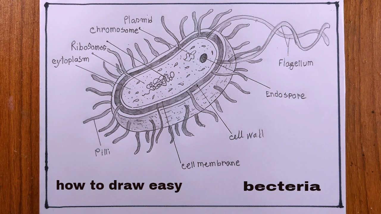
The cell wall also makes gram staining possible. Movement and chemotaxis are made possible by the motile organelle known as the flagellum. Web bacteria and archaea both have an organelle called the flagellum that resembles a filamentous thread. There are also cell walls, cytoplasm, and nucleoids. Download a free printable outline of this video and draw along with us.
Bacteria Drawing How To Draw Bacteria Step By Step
Web learn step by step drawing tutorial. Download a free printable outline of this video and draw along with us. Web how to draw bacteria.easy outline diagrambuy the material i used 1. The cell wall also makes gram staining possible. However, they can be as tiny as 0.3 µm and as large as 0.7mm.

How to Draw Bacteria Really Easy Drawing Tutorial
Outer layer (cell envelope), cell interior, and additional structures. Web thanks for watching, like, comment, share, and subscribe!how to draw bacteria#bacteria #stepbystep #easydrawing The size of these cells range between 1 micrometre to 3 micrometres, so they are barely visible under the light microscope. In this guide, we will be drawing 3 different shapes to represent the incredible variety on.
how to draw a bacteria easy/bacteria drawing YouTube
Download a free printable outline of this video and draw along with us. Web how to draw bacteria.easy outline diagrambuy the material i used 1. If you don't have a printer just keep this open. Web thanks for watching, like, comment, share, and subscribe!how to draw bacteria#bacteria #stepbystep #easydrawing However, they can be as tiny as 0.3 µm and as.
Bacteria Drawing How To Draw Bacteria Step By Step
The structure of the bacteria consists of three major parts: However, they can be as tiny as 0.3 µm and as large as 0.7mm. Scale 1:1 (1) how to draw: Web bacteria such as clostridium difficile can become problematic and is now a serious type of nosocomial infection. Free & simple drawing tutorials for kindergarten.
Web In Most Bacteria The Most Numerous Intracellular Structure Is The Ribosome, The Site Of Protein Synthesis In All Living Organisms.
They are also very versatile organisms, surviving in extremely inhospitable conditions. It is well organized and tightly packed, which explains its resistance to staining under the microscope. Larger bacterial cells may be visible. Download a free printable outline of this video and draw along with us.
Such Organisms Are Called Extremophiles.
The copy numbers of these proteins range from a few to. Web #bacteria #bacteriadiagram #adimushowbacteria is a prokaryotic cell. Web the structure of bacteria is known for its simple body design. The cell wall also makes gram staining possible.
The First One Will Be One Of The Easier Ones Of The Three To Ease You Into The Process!
This first one will be shaped a bit. Web in this video, we show you how to draw and label a basic bacterial cell. The limit of resolution with the unaided eye is about 200 microns, and as many bacteria are smaller than this size, they are not visible with naked eyes. Hence, they are classified as prokaryotic organisms.
We Use To See Them Via Microscope.
The size of these cells range between 1 micrometre to 3 micrometres, so they are barely visible under the light microscope. However, they can be as tiny as 0.3 µm and as large as 0.7mm. There are also cell walls, cytoplasm, and nucleoids. This means they do not have a nucleus or any other structures which are surrounded by.