Drawing Of The Uterus
Drawing Of The Uterus - Web the uterus is 6 to 8 cm (2.4 to 3.1 inches) long; Click to view large image. The vulva and its structures form the external genitalia. Web anatomy of the female reproductive system; This system of ducts connects to the ovaries, the primary reproductive organs. The endometrium (uterine mucous membrane) is lined with simple columnar epithelium (lamina epithelialis) and contains numerous tubular glands. The width of the organ varies; It's also called the womb. The video explains the structure of the. It is generally about 6 cm wide at the fundus and only half this distance at the isthmus.
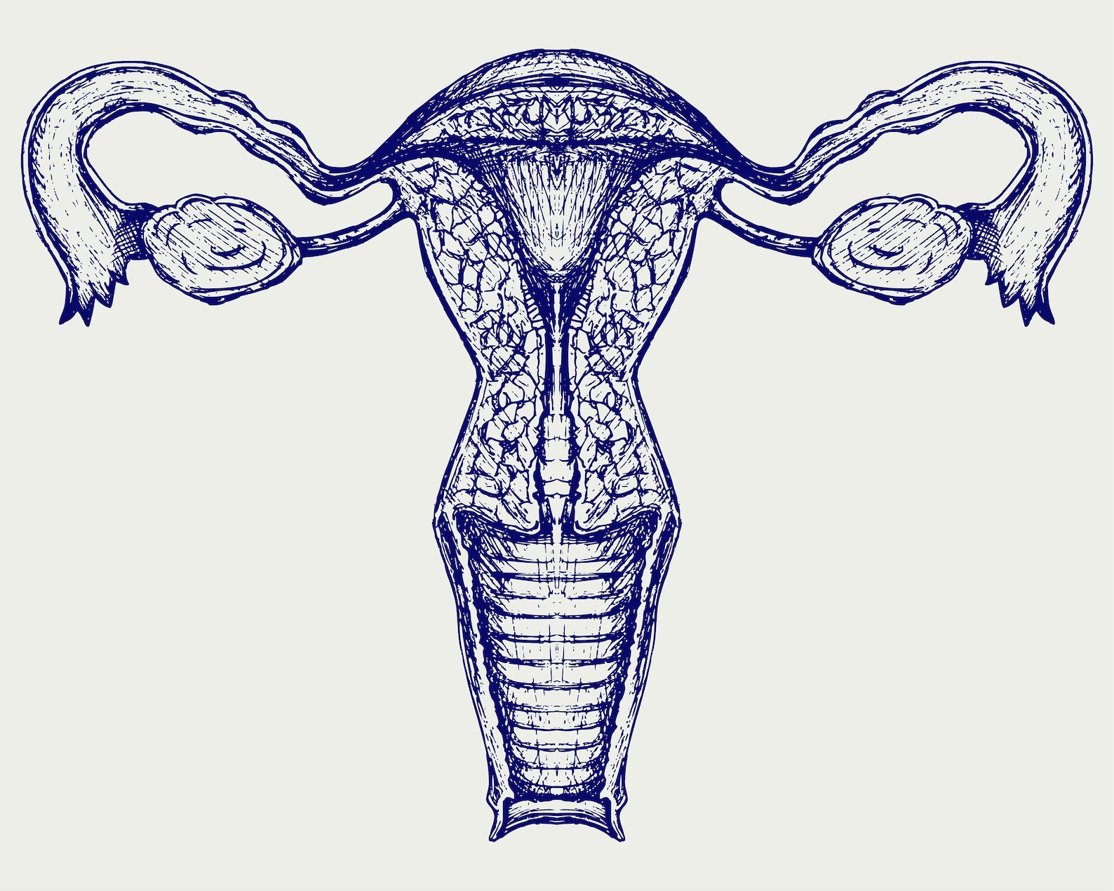
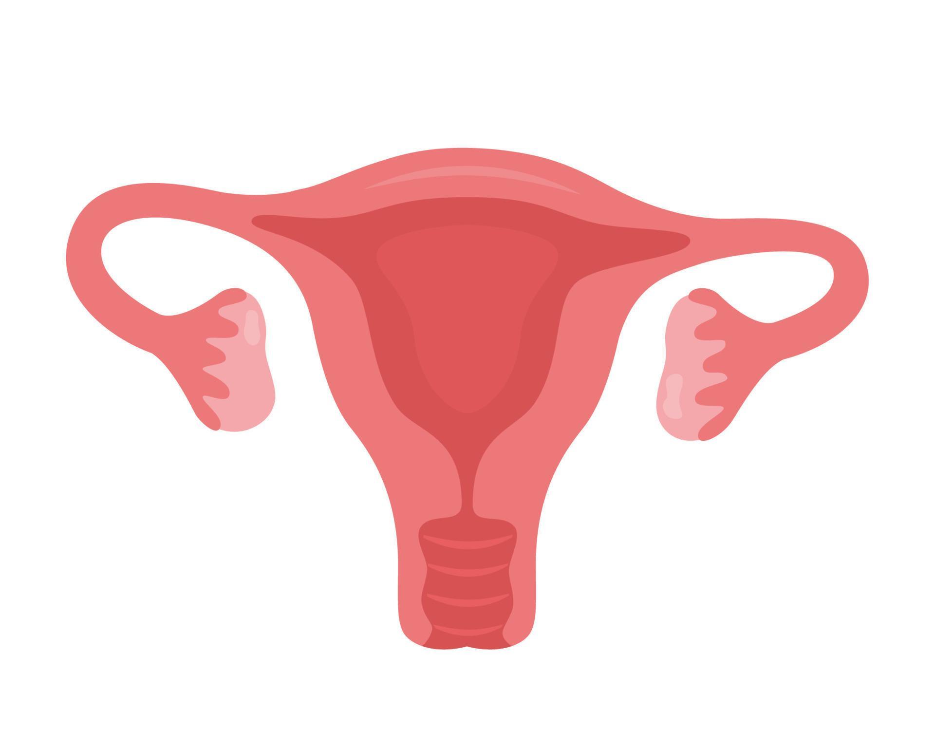
Web by kelly burch. Transformed a plain cloth line drawing of a uterus like this. Uteri) or womb ( / wuːm /) is the organ in the reproductive system of most female mammals, including humans, that accommodates the embryonic and fetal development of one or more embryos until birth. It is usually present in people assigned female at birth. Art nouveau design element for decoration drawing red flower 1899. The width of the organ varies; The uterus is located in the lower belly area between the hips (pelvis), through the vagina just past the cervix. This system of ducts connects to the ovaries, the primary reproductive organs. Mucosa (endometrium), muscularis ( myometrium) and serosa / adventitia ( perimetrium ). The uterus has three layers:
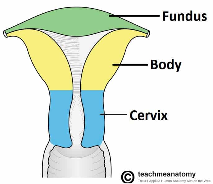
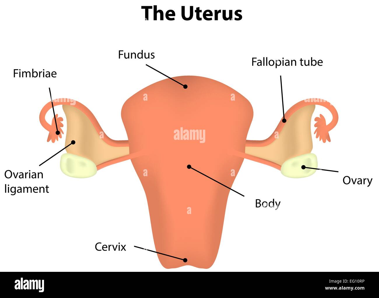
The uterus, also commonly known as the womb, is a hollow muscular organ of the female reproductive system that is responsible for the development of the embryo and fetus during pregnancy. Certain conditions and diseases of the uterus can cause painful symptoms that require medical treatment. A broad curved area in which fallopian tubes connect to the uterus, the corpus (body); The uterus is where a fetus develops during pregnancy. Web leonardo is credited with drawing the uterus with only one chamber, contradicting theories that the uterus was comprised of multiple chambers which many believed divided fetuses into separate compartments in the case of twins. The portion of the uterus superior to the opening of the uterine tubes is called the fundus. The female reproductive organs include several key structures, such as the ovaries, uterus, vagina, and vulva. The function of the uterine cervix during pregnancy is described at the end of the article. The uterus has three layers: Web histology of the uterus.
Internal and External Uterus Clipart Silhouette Clip Art Etsy
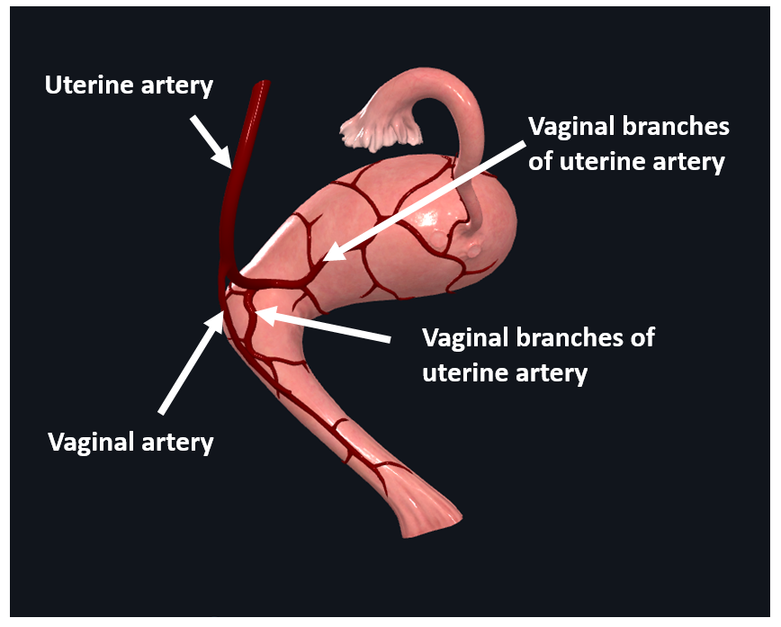
This short article describes the normal anatomy of the uterus and will focus on definitions, structure, location, supporting ligaments, blood supply and innervation. It is generally about 6 cm wide at the fundus and only half this distance at the isthmus. The uterus is where a fetus develops during pregnancy. A broad curved area in which fallopian tubes connect to.
The Uterus Structure Location Vasculature TeachMeAnatomy
The uterus, also known as the womb, is a hollow, muscular organ located in the pelvis between the bladder and rectum. Web the uterus has three parts; The uterus, also commonly known as the womb, is a hollow muscular organ of the female reproductive system that is responsible for the development of the embryo and fetus during pregnancy. Web the.

Uterus, Ovaries, Fallopian Tubes, Illustration Stock Image C043
Web the uterus (from latin uterus, pl.: It is generally about 6 cm wide at the fundus and only half this distance at the isthmus. Browse 668 uterus drawing photos and images available, or search for uterus illustration to find more great photos and pictures. The uterus, also commonly known as the womb, is a hollow muscular organ of the.

dibujo de arte de línea continua del útero reproductivo femenino
A lower neck region of the uterus, and. Web the uterus has three parts; The function of the uterine cervix during pregnancy is described at the end of the article. Human organs hand drawn line icon set. Web the uterus (from latin uterus, pl.:
Anatomy of the Uterus Female Reproductive Anatomy Geeky Medics
Web the uterus is a female secondary sex organ located within the pelvis. Human organs hand drawn line icon set. This short article describes the normal anatomy of the uterus and will focus on definitions, structure, location, supporting ligaments, blood supply and innervation. The uterus, also commonly known as the womb, is a hollow muscular organ of the female reproductive.
Uterus. Woman reproductive health illustration. Gynecology. Anatomy
Web the uterus is 6 to 8 cm (2.4 to 3.1 inches) long; Web uterine fibroids can cause anemia and fatigue with heavy or long menstrual periods. A lower neck region of the uterus, and. These fully annotated anatomical illustrations are presented as a comprehensive atlas of the. Web the uterus (from latin uterus, pl.:
Uterus Labeled Diagram Stock Vector Image & Art Alamy
This system of ducts connects to the ovaries, the primary reproductive organs. Its average size is approximately 5 cm wide by 7 cm long (approximately 2 in by 3 in) when a female is not pregnant. Certain conditions and diseases of the uterus can cause painful symptoms that require medical treatment. A broad curved area in which fallopian tubes connect.

Uterus Anatomy Worksheet Single FILLED Digital Download Human Anatomy
It’s hollow and muscular and sits between your rectum and bladder in your pelvis. A lower neck region of the uterus, and. The main part of a uterus, and it starts directly below the level of fallopian tubes and continues downward, isthmus; Transformed a plain cloth line drawing of a uterus like this. The vulva and its structures form the.
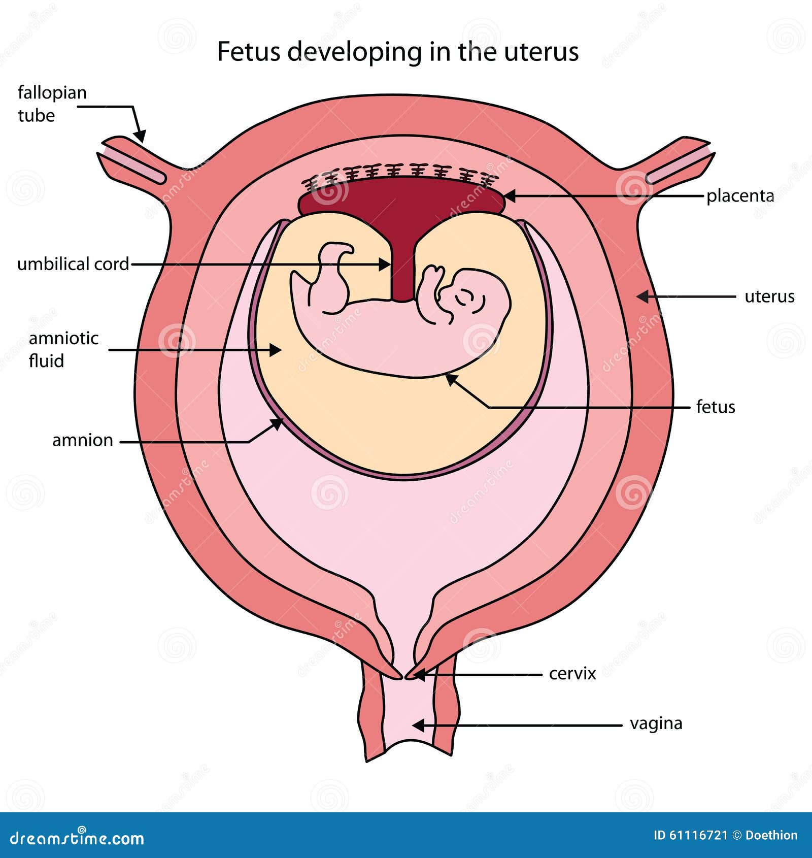
Diagram Of Uterus With Labels
Human organs hand drawn line icon set. The endometrium (uterine mucous membrane) is lined with simple columnar epithelium (lamina epithelialis) and contains numerous tubular glands. It is generally about 6 cm wide at the fundus and only half this distance at the isthmus. Web the uterus is the muscular organ that nourishes and supports the growing embryo (see figure 27.14)..
Vector Isolated Illustration of Uterus Stock Vector Illustration of
It’s hollow and muscular and sits between your rectum and bladder in your pelvis. Web the female reproductive system includes external and internal genitalia. The main part of a uterus, and it starts directly below the level of fallopian tubes and continues downward, isthmus; The function of the uterine cervix during pregnancy is described at the end of the article..
Uteri) Or Womb ( / Wuːm /) Is The Organ In The Reproductive System Of Most Female Mammals, Including Humans, That Accommodates The Embryonic And Fetal Development Of One Or More Embryos Until Birth.
The function of the uterine cervix during pregnancy is described at the end of the article. Web uterine fibroids can cause anemia and fatigue with heavy or long menstrual periods. Drawing shows the uterus, myometrium (muscular outer layer of the uterus), endometrium (inner lining of the uterus), ovaries, fallopian tubes, cervix, and vagina. Human organs hand drawn line icon set.
Into Beautiful Works Of Art Like These.
This short article describes the normal anatomy of the uterus and will focus on definitions, structure, location, supporting ligaments, blood supply and innervation. It’s hollow and muscular and sits between your rectum and bladder in your pelvis. The uterine cavity opens into the vaginal cavity, and the two make up what is commonly known as the birth canal. The uterus has three layers of muscle and is one of the strongest muscles in the body.
The Female Reproductive Organs Include Several Key Structures, Such As The Ovaries, Uterus, Vagina, And Vulva.
Human organs hand drawn line icon set. The uterine tubes, the uterus, and the vagina. The uterus is where a fetus develops during pregnancy. This part is structurally and functionally different to the rest of the uterus.
The Uterus Is Located In The Lower Belly Area Between The Hips (Pelvis), Through The Vagina Just Past The Cervix.
It's also called the womb. This system of ducts connects to the ovaries, the primary reproductive organs. A broad curved area in which fallopian tubes connect to the uterus, the corpus (body); The vulva and its structures form the external genitalia.