Embryology Drawings
Embryology Drawings - He also dramatically reformed the field of human embryology. Haeckel s famous drawings are a creationist cause c l bre (3). However, the assertion by explore evolution that haeckel claimed that top row represented earliest embryos is false. Data from embryology are fully consistent with darwinian evolution. This version obscures the differences between the earliest stages of embryos as egregiously as haeckel’s original drawings did. Web the theory of recapitulation, also called the biogenetic law or embryological parallelism —often expressed using ernst haeckel 's phrase ontogeny recapitulates phylogeny —is an historical hypothesis that the development of the embryo of an animal, from fertilization to gestation or hatching ( ontogeny ), goes through stages resembling or. Later, in everyday biology (curtis et al. One of the most controversial drawings in evolutionary biology. Web a recent study (1) coauthored by several of us and discussed by elizabeth pennisi (research news, 5 sept. Yet, haeckel's embryo grids are much more complex than any textbook explanation.
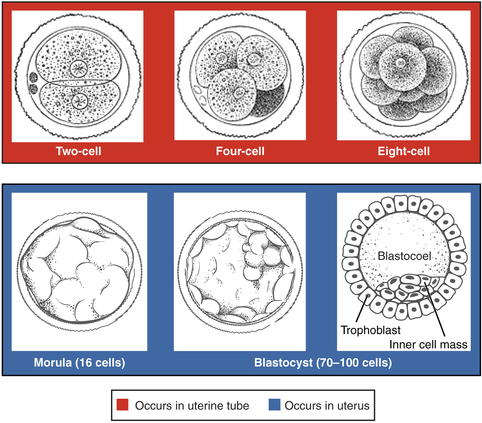
Web embryo drawing is the illustration of embryos in their developmental sequence. Web the sixth embryo set in the series, haeckel’s calf (rind in german) became a sheep in gruenberg’s version. Web pictures from the past powerfully shape current views of the world. I n the early stages of human embryonic development, a zygote divides into two identical totipotent. Web embryo drawing is the illustration of embryos in their developmental sequence. However, the assertion by explore evolution that haeckel claimed that top row represented earliest embryos is false. More than a century ago, ernst haeckel created embryo drawings to illustrate the morphological similarity of vertebrate early embryos. This is haeckel's embryo grid, the most common of all illustrations in biology textbooks. Web embryo drawings drawn by haeckel in 1866 for his recapitulation theory. He recaptures the shocking novelty of pictures that enthralled schoolchildren.
Web these drawings have been both widely presented and frequently criticized. Web in haeckel’s embryos: Images, evolution and fraud, published by the university of chicago press, dr nick hopwood tells the full story for the first time. This version obscures the differences between the earliest stages of embryos as egregiously as haeckel’s original drawings did. In plants and animals, an embryo develops from a zygote, the single cell that results when an egg and sperm fuse during fertilization. As we discover in haeckel’s embryos, german biologist ernst haeckel included illustrations of the embryological stages of vertebrates in a series of books published between 1868. Web embryo drawings drawn by haeckel in 1866 for his recapitulation theory. Web a recent study (1) coauthored by several of us and discussed by elizabeth pennisi (research news, 5 sept. Haeckel s famous drawings are a creationist cause c l bre (3). Haeckel represented this idea with drawings of vertebrate embryos at similar developmental stages.

Human embryo Royalty Free Vector Image VectorStock
Web haeckel represented this idea with drawings of vertebrate embryos at similar developmental stages. Later, in everyday biology (curtis et al. Images, evolution and fraud, published by the university of chicago press, dr nick hopwood tells the full story for the first time. In books, television programs, and websites, new images appear alongside others that have survived from decades ago..

Embryo Development A Development process of Fetus Week by Week
In animals, the zygote divides repeatedly to form a ball of cells, which then forms a set of tissue layers that migrate and fold to. Web ernst haeckel and comparative embryology. This is haeckel's embryo grid, the most common of all illustrations in biology textbooks. This is haeckel’s embryo grid, the most common of all illustrations in biology textbooks. Among.
Embryonic Development · Anatomy and Physiology
Haeckel s famous drawings are a creationist cause c l bre (3). Web accuracy in embryo illustrations. Web in haeckel’s embryos: A team of researchers labeled one of two cells in a developing embryo with gfp and used dna (blue) and actin (pink) labeling to track cell progeny to determine the contribution of each to developing structures. He also dramatically.
Embryonic Development · Anatomy and Physiology
Web embryo drawing is the illustration of embryos in their developmental sequence. This is haeckel's embryo grid, the most common of all illustrations in biology textbooks. This is ernst haeckel's own drawing of a series of embryos, from the german first edition of 'anthropogenie', 1874, in original sepia/gold lithography. As we discover in haeckel’s embryos, german biologist ernst haeckel included.
Stages human embryonic development Royalty Free Vector Image
Among the most famous are drawings of embryos by the darwinist ernst haeckel in which humans and other vertebrates begin identical, then diverge toward their. Web haeckel believed that the development of an embryo revealed the adult stages of the organism's ancestors. Web haeckel represented this idea with drawings of vertebrate embryos at similar developmental stages. Our work has been.
Human embryonic development in human infographic 6158571 Vector Art at
This is haeckel's embryo grid, the most common of all illustrations in biology textbooks. This is ernst haeckel's own drawing of a series of embryos, from the german first edition of 'anthropogenie', 1874, in original sepia/gold lithography. The faithful gruenberg variation represented only the second appearance of haeckel’s full embryo set in a high school text. Web embryo drawing is.

What is an Embryo? (with pictures)
Web haeckel believed that the development of an embryo revealed the adult stages of the organism’s ancestors. He is most well known for his descriptions of phylogenetic trees, studies of radiolarians, and illustrations of vertebrate embryos to support his biogenetic law and darwin’s work with evolution. Among the most famous are drawings of embryos by the darwinist ernst haeckel in.

Stages in human embryonic development Royalty Free Vector
He discovered, described and named thousands of new species, mapped a genealogical tree relating all life. This is haeckel’s embryo grid, the most common of all illustrations in biology textbooks. The faithful gruenberg variation represented only the second appearance of haeckel’s full embryo set in a high school text. Images, evolution and fraud, published by the university of chicago press,.
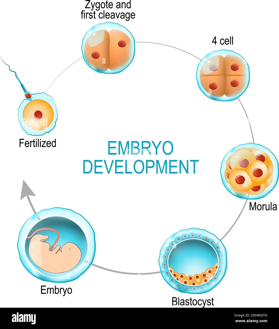
embryo development. from fertilization to zygote, morula and Blastocyst
Web the sixth embryo set in the series, haeckel’s calf (rind in german) became a sheep in gruenberg’s version. 1435) examined inaccuracies in embryo drawings published last century by ernst haeckel. Web pictures from the past powerfully shape current views of the world. Later, in everyday biology (curtis et al. (1) as seen here, the textbook uses a colorized and.

Paper Description of a 4 mm human embryo (1906) Embryology
Haeckel represented this idea with drawings of vertebrate embryos at similar developmental stages. Yet, haeckel's embryo grids are much more complex than any textbook explanation. He also dramatically reformed the field of human embryology. However, the assertion by explore evolution that haeckel claimed that top row represented earliest embryos is false. Web haeckel believed that the development of an embryo.
This Is Ernst Haeckel's Own Drawing Of A Series Of Embryos, From The German First Edition Of 'Anthropogenie', 1874, In Original Sepia/Gold Lithography.
Web and finally, as someone who studied developmental biology, i still find the neat little drawings to be utterly entrancing. Web ernst haeckel and comparative embryology. Among the most famous are drawings of embryos by the darwinist ernst haeckel in which humans and other vertebrates begin identical, then diverge toward their. He is most well known for his descriptions of phylogenetic trees, studies of radiolarians, and illustrations of vertebrate embryos to support his biogenetic law and darwin’s work with evolution.
Web In Haeckel’s Embryos:
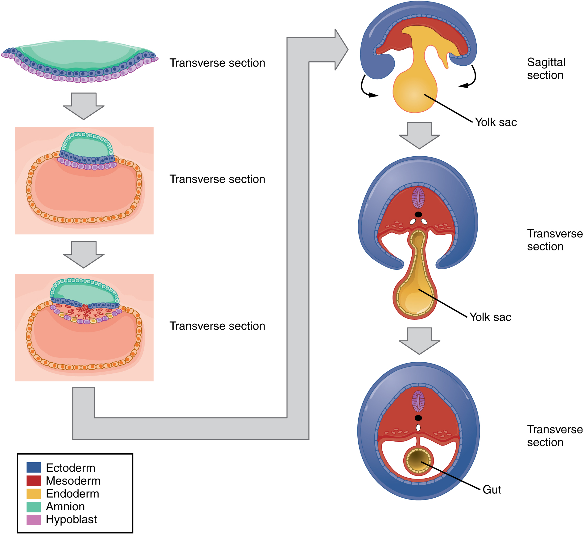
Our work has been used in a nationally televised debate to attack evolutionary theory, and to suggest that evolution cannot. Web these drawings have been both widely presented and frequently criticized. In animals, the zygote divides repeatedly to form a ball of cells, which then forms a set of tissue layers that migrate and fold to form an early embryo. Web embryo drawing is the illustration of embryos in their developmental sequence.
Web Embryo Drawings Drawn By Haeckel In 1866 For His Recapitulation Theory.
Yet, haeckel's embryo grids are much more complex than any textbook explanation. You cannot overwrite this file. Web the sixth embryo set in the series, haeckel’s calf (rind in german) became a sheep in gruenberg’s version. A team of researchers labeled one of two cells in a developing embryo with gfp and used dna (blue) and actin (pink) labeling to track cell progeny to determine the contribution of each to developing structures.
Web Haeckel Believed That The Development Of An Embryo Revealed The Adult Stages Of The Organism’s Ancestors.
Web however, as richardson and colleagues note, this hardly undermines the strong support for common descent from embryology, despite the claims of creationists and id proponents. Web the leading anatomist wilhelm his became a bitter enemy of haeckel and disputed the veracity of his embryo drawings. He also dramatically reformed the field of human embryology. In books, television programs, and websites, new images appear alongside others that have survived from decades ago.