Heart Anatomy Draw
Heart Anatomy Draw - The valves of the heart. Your brain and nervous system direct your heart’s function. Use a pen or pencil to draw the heart's main body. Right, left, superior, and inferior: Part of the teachme series. The middle layer is the myocardium. Web the slight deviation of the apex to the left is reflected in a depression in the medial surface of the inferior lobe of the left lung, called the cardiac notch. Create a curved shape similar to an acorn or apple’s bottom half. Angle the slightly tampered end. The position of the heart in the torso between the vertebrae and sternum (see figure 19.2 for the position of the heart within the thorax) allows for individuals to apply an emergency technique known as cardiopulmonary resuscitation (cpr) if the heart of a patient should stop.
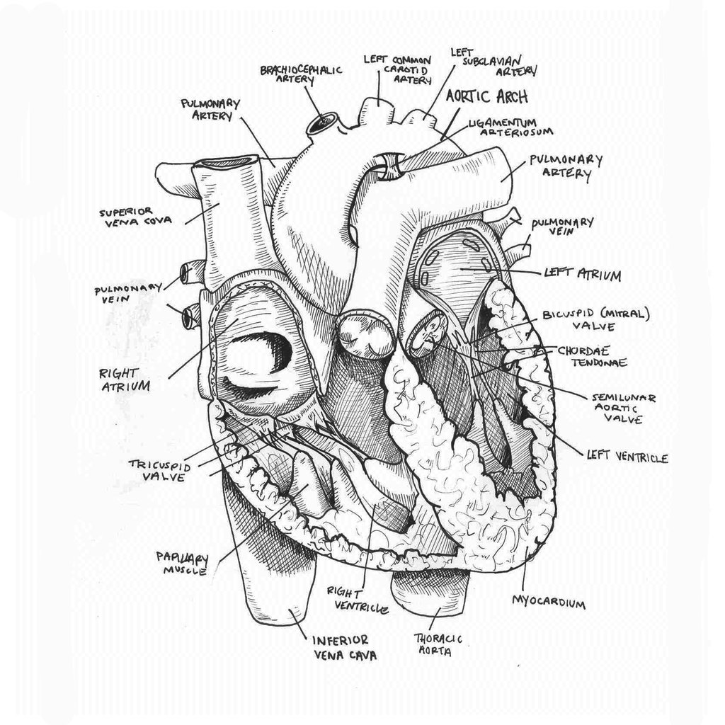
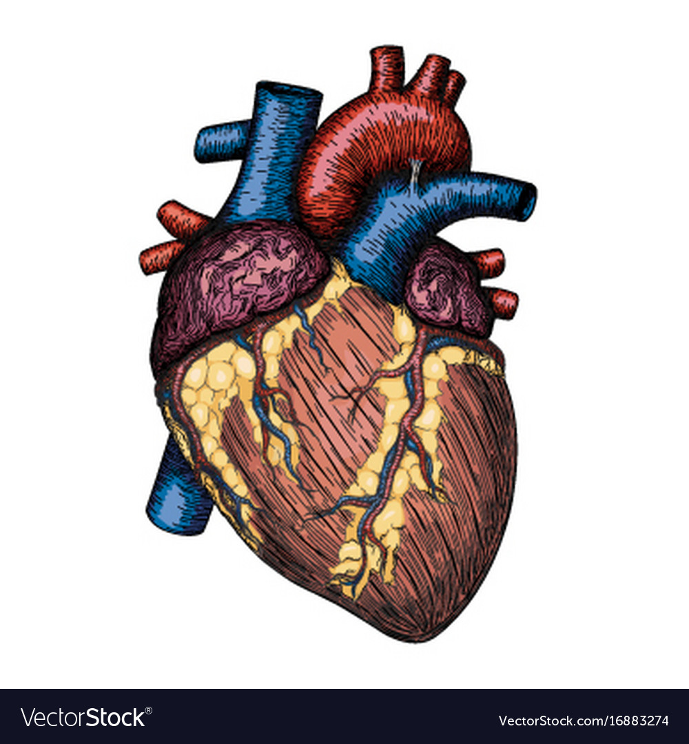
The muscular wall of the heart has three layers. Web worksheet showing unlabelled heart diagrams. Heart anatomy drawing stock photos are available in a variety of sizes and formats to fit your needs. Your brain and nervous system direct your heart’s function. Location of heart in the thorax. A drawing of the anatomy of the opened normal heart, with english labels. This is the superior vena cava. Web the slight deviation of the apex to the left is reflected in a depression in the medial surface of the inferior lobe of the left lung, called the cardiac notch. Web the surfaces and borders of the heart. In fishes the heart is a folded tube, with three or four enlarged areas that.
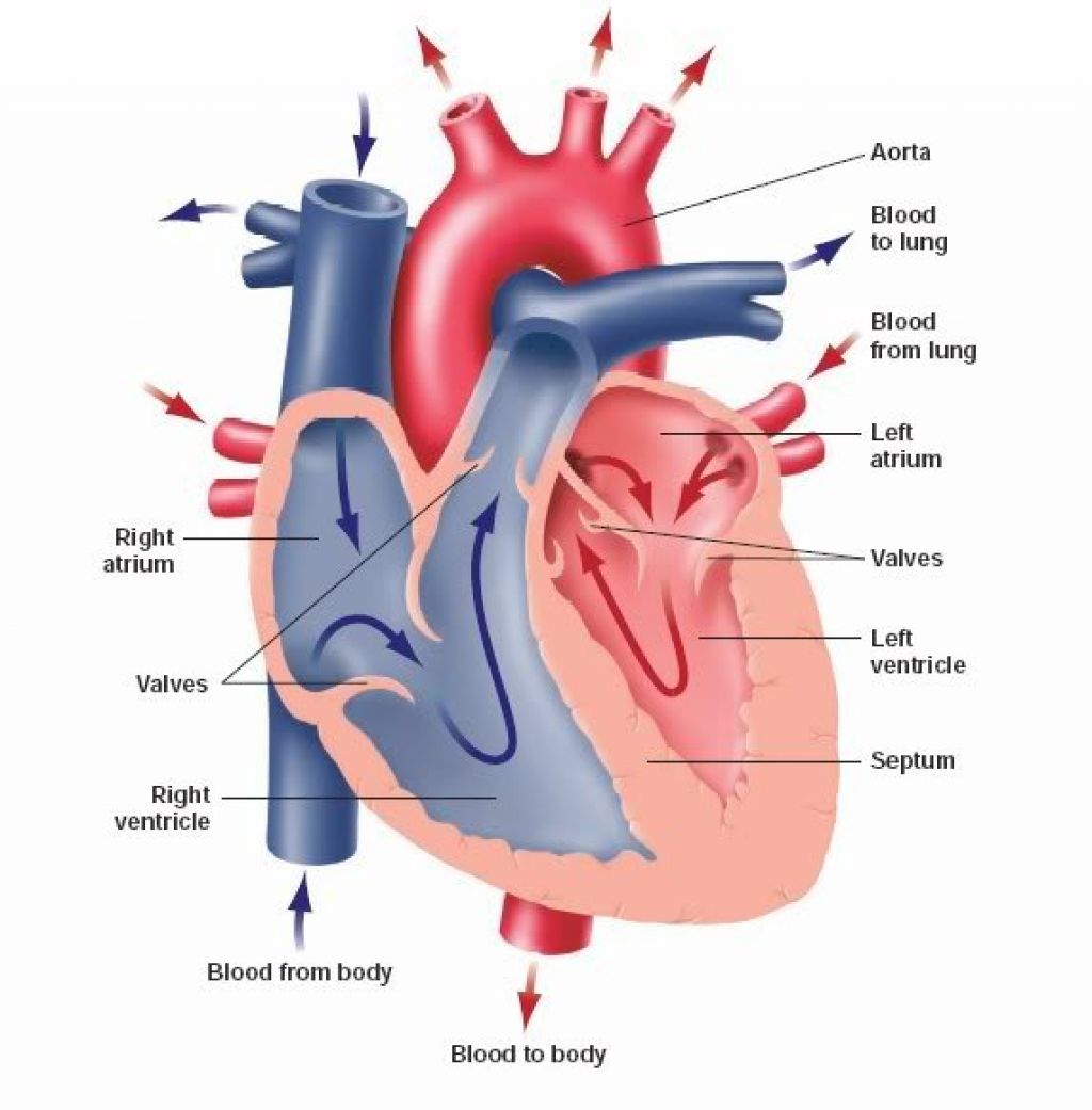
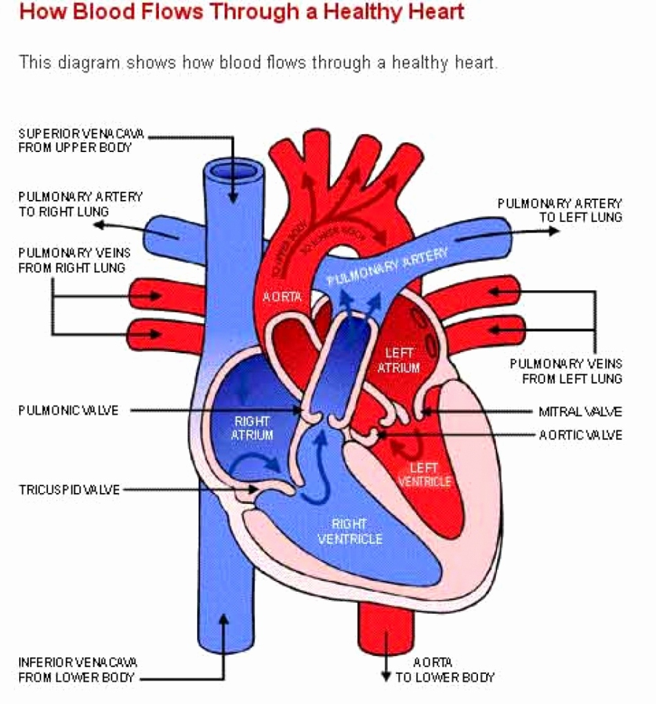
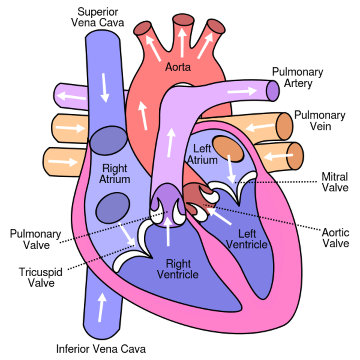
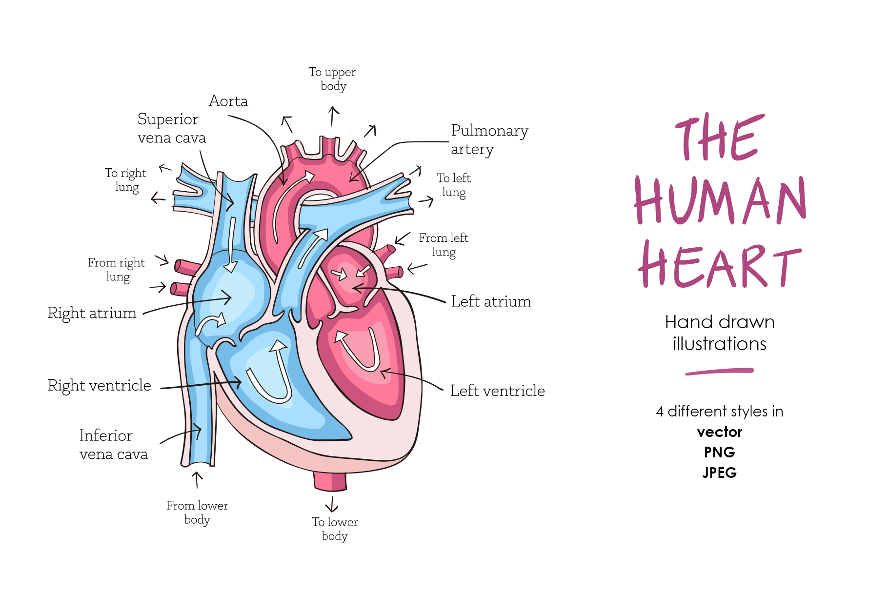
Web anatomy of the heart made easy along with the blood flow through the cardiac structures, valves, atria, and ventricles. Web function and anatomy of the heart made easy using labeled diagrams of cardiac structures and blood flow through the atria, ventricles, valves, aorta, pulmonary arteries veins, superior inferior vena cava, and chambers. This is the superior vena cava. Next, draw the right atrium and the superior vena cava. From the openstax anatomy and physiology book. Heart anatomy drawing stock photos are available in a variety of sizes and formats to fit your needs. Start with the pulmonary veins. The valves of the heart. Web muscle and tissue make up this powerhouse organ. Web heart, organ that serves as a pump to circulate the blood.
Anatomical Drawing Heart at GetDrawings Free download
Use a pen or pencil to draw the heart's main body. The outermost layer is the epicardium (or visceral pericardium). Electrical impulses make your heart beat, moving blood through these chambers. For the atrium, enclose an irregular form at the junction of the ventricle and the aortic arch. Web the heart has three layers.

How to Draw the Internal Structure of the Heart 13 Steps
The epicardium covers the heart, wraps around the roots of the great blood vessels, and adheres the heart wall to a protective sac. Start with the pulmonary veins. Web in this lecture, dr mike shows the two best ways to draw and label the heart! From the openstax anatomy and physiology book. The outermost layer is the epicardium (or visceral.
When one teaches, two learn. The heart and the circulatory system
This will make the pen drawing process much easier as we use the pencil marks for guidance. Use a pen or pencil to draw the heart's main body. Find a piece of paper and something to draw with. It may be a straight tube, as in spiders and annelid worms, or a somewhat more elaborate structure with one or more.
Human Heart Drawing Simple at Explore collection
Web the slight deviation of the apex to the left is reflected in a depression in the medial surface of the inferior lobe of the left lung, called the cardiac notch. Web the heart has three layers. Web heart drawing realistic anatomical beauty: Web muscle and tissue make up this powerhouse organ. Create a curved shape similar to an acorn.
Learn About the Heart and Circulatory System for Kids HubPages
By applying pressure with the flat portion of one hand on the. Heart anatomy drawing stock photos are available in a variety of sizes and formats to fit your needs. Web drawing the heart anatomy can be challenging, but with practice and dedication, it can become easier over time! The outermost layer is the epicardium (or visceral pericardium). This thick.
Human heart hand drawn anatomical sketch Vector Image
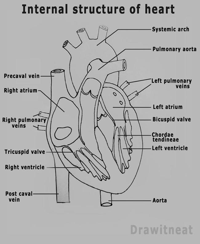
A drawing of the anatomy of the opened normal heart, with english labels. Web anatomy of the heart: Right, left, superior, and inferior: Electrical impulses make your heart beat, moving blood through these chambers. Web worksheet showing unlabelled heart diagrams.
Human heart anatomy (274491) Illustrations Design Bundles
Web to draw an anatomical heart requires a lot of attention and patience. Drawing your boyfriend a cute emoji, maybe the kissing emoji, the smiley emoji or. It may be a straight tube, as in spiders and annelid worms, or a somewhat more elaborate structure with one or more receiving chambers (atria) and a main pumping chamber (ventricle), as in.

How to Draw the Internal Structure of the Heart (with Pictures)
The position of the heart in the torso between the vertebrae and sternum (see figure 19.2 for the position of the heart within the thorax) allows for individuals to apply an emergency technique known as cardiopulmonary resuscitation (cpr) if the heart of a patient should stop. This thick layer is the muscle that contracts to pump and propel blood. Use.
DRAW IT NEAT How to draw human heart labeled
Next, draw the right atrium and the superior vena cava. It also has several margins: This interactive atlas of human heart anatomy is based on medical illustrations and cadaver photography. The medical information on this site is provided as an information resource only, and is not to be used or relied on for any. Xxxl very detailed human heart.

Heart Anatomy chambers, valves and vessels Anatomy & Physiology
Use a pen or pencil to draw the heart's main body. The position of the heart in the torso between the vertebrae and sternum (see figure 19.2 for the position of the heart within the thorax) allows for individuals to apply an emergency technique known as cardiopulmonary resuscitation (cpr) if the heart of a patient should stop. The heart has.
Angle The Slightly Tampered End.
Heart anatomy drawing stock photos are available in a variety of sizes and formats to fit your needs. Create a curved shape similar to an acorn or apple’s bottom half. The heart wall is composed of three layers. This interactive atlas of human heart anatomy is based on medical illustrations and cadaver photography.
A Drawing Of The Anatomy Of The Opened Normal Heart, With English Labels.
Your brain and nervous system direct your heart’s function. Find a piece of paper and something to draw with. Web old chromolithograph illustration of human vital organs. Part of the teachme series.
Base (Posterior), Diaphragmatic (Inferior), Sternocostal (Anterior), And Left And Right Pulmonary Surfaces.
For the atrium, enclose an irregular form at the junction of the ventricle and the aortic arch. Web to draw an anatomical heart requires a lot of attention and patience. Web in this lecture, dr mike shows the two best ways to draw and label the heart! Web function and anatomy of the heart made easy using labeled diagrams of cardiac structures and blood flow through the atria, ventricles, valves, aorta, pulmonary arteries veins, superior inferior vena cava, and chambers.
It Also Has Several Margins:
Next, draw the right atrium and the superior vena cava. Xxxl very detailed human heart. Web the heart has three layers. The muscular wall of the heart has three layers.