Histology Drawing
Histology Drawing - Web histology is that it is the structural basis for cell, tissue and organ biology and function (physiology) and disease (pathology). Connective tissue including cartilage and bone blood and blood formation 3. Web draw a schematic representation of the passage of urine from the kidneys to the urethra. Web 12/05/2023 30/04/2021 by sonnet poddar. Web they are sketches from selected slides used in class from the teaching slide set. Get expert guidance and personalized support from a professor. The liver performs several important functions in the human body, such as given below: A simple squamous epithelium is a single layer of flat cells in. There are no labeled images here, as the goal is to get you to practice identifying structures on your own. These labelled diagrams should closely follow the current science courses in histology, anatomy and embryology and complement the virtual microscopy used in the current medical course.
The histology laboratory drawings resource contains 104 hand drawn sketches by dr. Web histology is that it is the structural basis for cell, tissue and organ biology and function (physiology) and disease (pathology). Both surface and side view has been demonstrated in this video. Connective tissue including cartilage and bone blood and blood formation 3. In this part of the article i am going to show you the most important histological features from a esophagus histology. Web draw a schematic representation of the passage of urine from the kidneys to the urethra. Get expert guidance and personalized support from a professor. As a library, nlm provides access to scientific literature. Web mucosa of esophagus structure. Kidney (236) examine this slide with your naked eye or at minimum zoom.
Histology is organized into four basic types of tissues. Web draw a schematic representation of the passage of urine from the kidneys to the urethra. Web in the next tutorial, our demonstrator veronika stankova shares with you examples and tips on how to draw histological diagrams from templates. Web histology is that it is the structural basis for cell, tissue and organ biology and function (physiology) and disease (pathology). In the parenchyma of spleen histology slide, you will find the red pulp and white pulp. These labelled diagrams should closely follow the current science courses in histology, anatomy and embryology and complement the virtual microscopy used in the current medical course. Christensen for the laboratory sessions he conducted in the medical histology course for first year medical students. Liver histology drawing, step by step drawing of histology of liver. The liver performs several important functions in the human body, such as given below: Web it examines the correlation between structure and function.

Histology Drawing of Loose Areolar Tissue with explanation connective
The thyroid gland is situated opposite c5 and t1 vertebra in the anterior neck region. There are no labeled images here, as the goal is to get you to practice identifying structures on your own. J taibah univ med sci. You are responsible for 25 drawings*. Cardiac muscle shows many structural and functional characteristics intermediate between skeletal and smooth muscles.
Histology illustration A4 art print cell drawing medical Etsy
Connective tissue including cartilage and bone blood and blood formation 3. 2.8k views 3 years ago. In the parenchyma of spleen histology slide, you will find the red pulp and white pulp. Dr carol lazer, april 2005. Christensen for the laboratory sessions he conducted in the medical histology course for first year medical students.

Basic Histology Diagrams in Colour ANAT2241 Histology Basic and
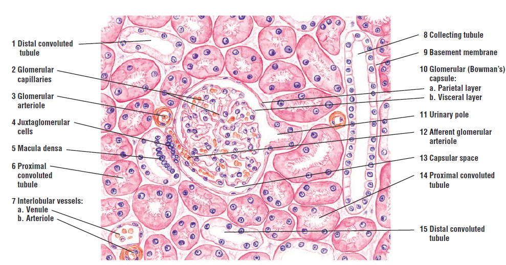
Each should include all distinctive/characteristic features clearly labeled. A simple squamous epithelium is a single layer of flat cells in. What happens in each part of the kidney? Connective tissue including cartilage and bone blood and blood formation 3. Get expert guidance and personalized support from a professor.

Histology Fundamentals Ovarian Follicle Histology Draw It to Know It
Organs are assembled from the four basic types of tissues and have cells with specialized functions. Kidney (236) examine this slide with your naked eye or at minimum zoom. J taibah univ med sci. Both surface and side view has been demonstrated in this video. You are responsible for 25 drawings*.

Liver histology Drawing YouTube
J taibah univ med sci. Web these lab worksheets are designed to guide you through a laboratory experience in histology. What happens in each part of the kidney? Christensen for the laboratory sessions he conducted in the medical histology course for first year medical students. Web 25/04/2023 16/07/2021 by sonnet poddar.

Histology Drawings February 2014
Tunica serosa or adventitia of esophagus. Before going to details description of spleen structure i would like to enlist the important structures that you might identify under light microscope. Web bulleted notes & final drawings. As a library, nlm provides access to scientific literature. You are responsible for 25 drawings*.

Histology Drawings January 2014
Web dr yogesh ganorkar tells about how to draw histological diagram of large intestine. Cells are the tiny living units that form the tissues, organs and structures within the body. Web liver histology drawing. Dr carol lazer, april 2005. Christensen for the laboratory sessions he conducted in the medical histology course for first year medical students.

Histology Drawings Urinary System
Web bulleted notes & final drawings. Get expert guidance and personalized support from a professor. You will find some salient microscopic features of each region of the small intestine. Web liver histology drawing. The histology laboratory drawings resource contains 104 hand drawn sketches by dr.
Histology Drawings
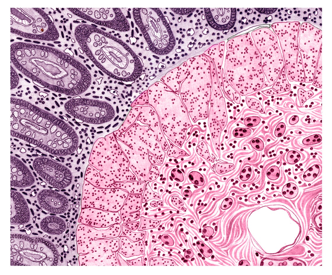
Web dr yogesh ganorkar tells about how to draw histological diagram of large intestine. Tunica serosa or adventitia of esophagus. Web mucosa of esophagus structure. In this part of the article i am going to show you the most important histological features from a esophagus histology. In this short article, i will discuss duodenum histology in detail, along with its.

Histology Drawings January 2014
The first pages illustrate introductory concepts for those new to microscopy as well as definitions of commonly used histology terms. Dr carol lazer, april 2005. Each should include all distinctive/characteristic features clearly labeled. Kidney (236) examine this slide with your naked eye or at minimum zoom. Web histology is the microscopic study of tissues and cells used in understanding the.
Before Going To Details Description Of Spleen Structure I Would Like To Enlist The Important Structures That You Might Identify Under Light Microscope.
Dr carol lazer, april 2005. Web in the next tutorial, our demonstrator veronika stankova shares with you examples and tips on how to draw histological diagrams from templates. In this part of the article i am going to show you the most important histological features from a esophagus histology. Web 12/05/2023 30/04/2021 by sonnet poddar.
Web 25/04/2023 16/07/2021 By Sonnet Poddar.
Today, in this short article, i will show you the important histological features from the cardiac muscle histology slide. Web histology is the microscopic study of tissues and cells used in understanding the pathogenesis and diagnosis of various diseases. Web bulleted notes & final drawings. Kidney (236) examine this slide with your naked eye or at minimum zoom.
The Liver Performs Several Important Functions In The Human Body, Such As Given Below:
Web draw a schematic representation of the passage of urine from the kidneys to the urethra. Web histology diagram of simple squamous epithelium histology diagram. The histology laboratory drawings resource contains 104 hand drawn sketches by dr. The first pages illustrate introductory concepts for those new to microscopy as well as definitions of commonly used histology terms.
What Is The Plan For The Study Of Cells, Tissues And Organs?
Cardiac muscle shows many structural and functional characteristics intermediate between skeletal and smooth muscles. Web spleen histology drawing. You are responsible for 25 drawings*. These labelled diagrams should closely follow the current science courses in histology, anatomy and embryology and complement the virtual microscopy used in the current medical course.