How To Draw A Ribosome
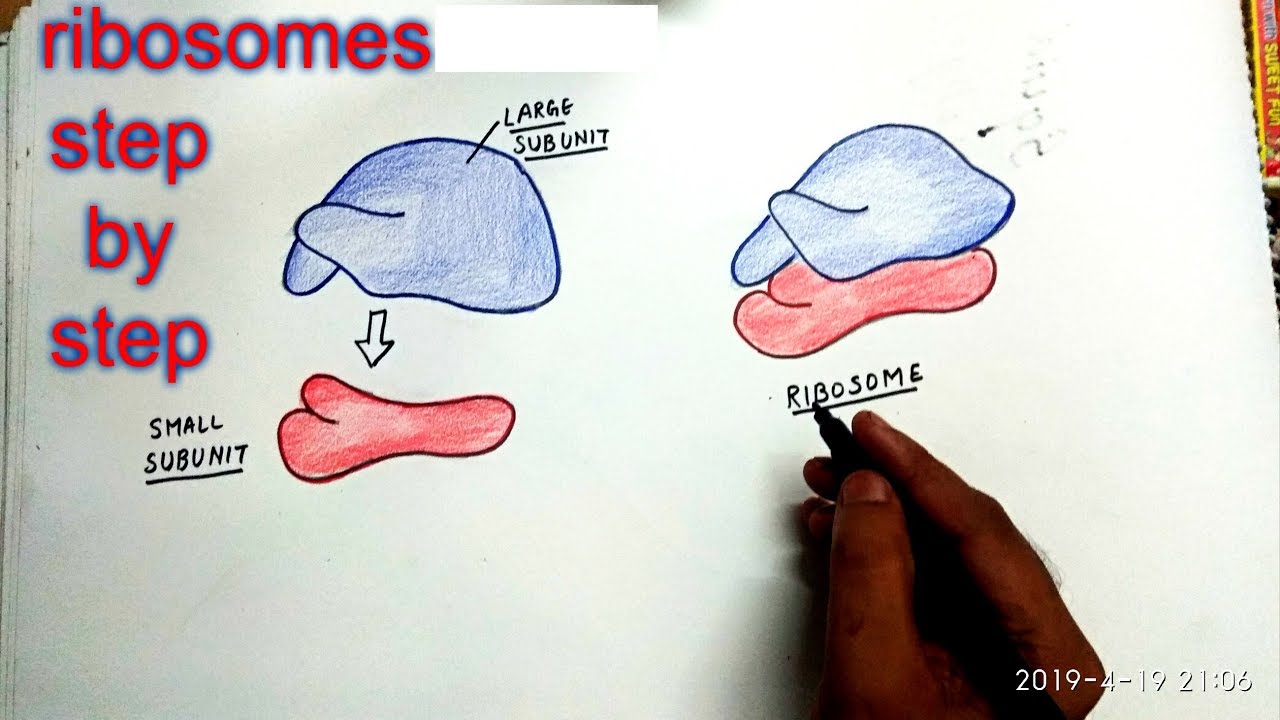
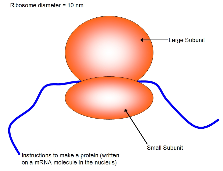
How To Draw A Ribosome - Coli, there are between 10,000 and 70,000 ribosomes present in each cell at any given time.a ribosome is a complex macromolecule composed of structural and catalytic rrnas, and many distinct polypeptides. A simple method for ribosome drawing. This chain of amino acids then folds to form a complex 3d structure. The translation of information and the linking of amino acids are at the heart of the protein production process.a ribosome, formed from two subunits locking together, functions to: The large subunit sits on top of the small subunit, with an rna template sandwiched between the two. Sure enough, they found that ribosomes that build the pol protein stopped just before the very end, leaving the protein tethered to the ribosome like a balloon tied to a child’s hand. As catalysts they speed the time of reactions, as fibers they provide support, and many proteins function in specific tasks, like contracting muscle cells. Web figure 3.4.1 3.4. They are also found in the mitochondria and chloroplast of a. Web this tutorial demonstrates how to draw ribosomes in powerpoint for research publication, conference posters, science figures and graphical abstracts.
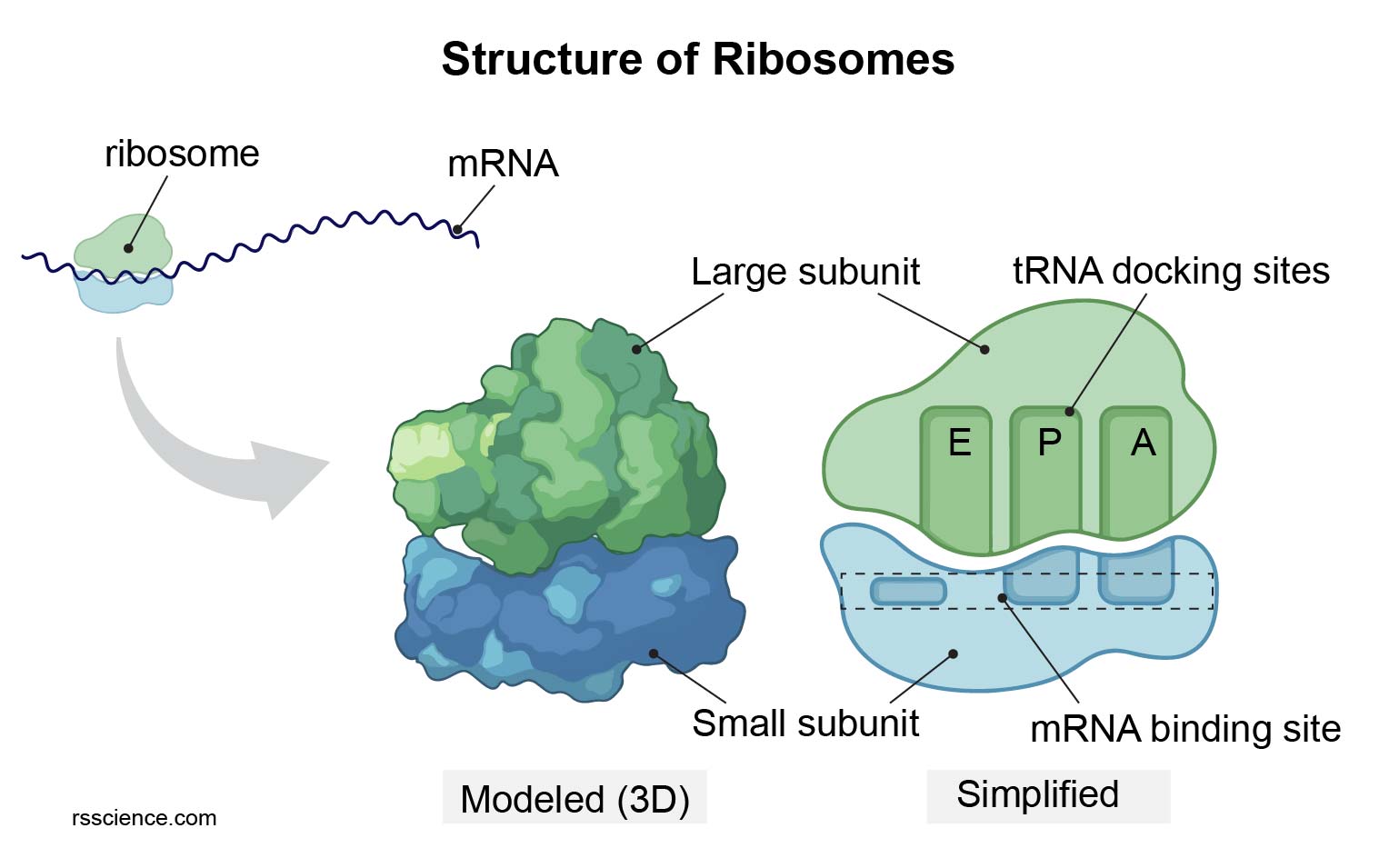
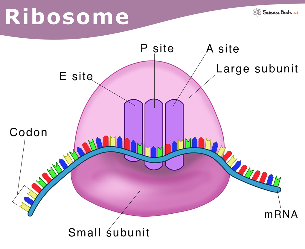
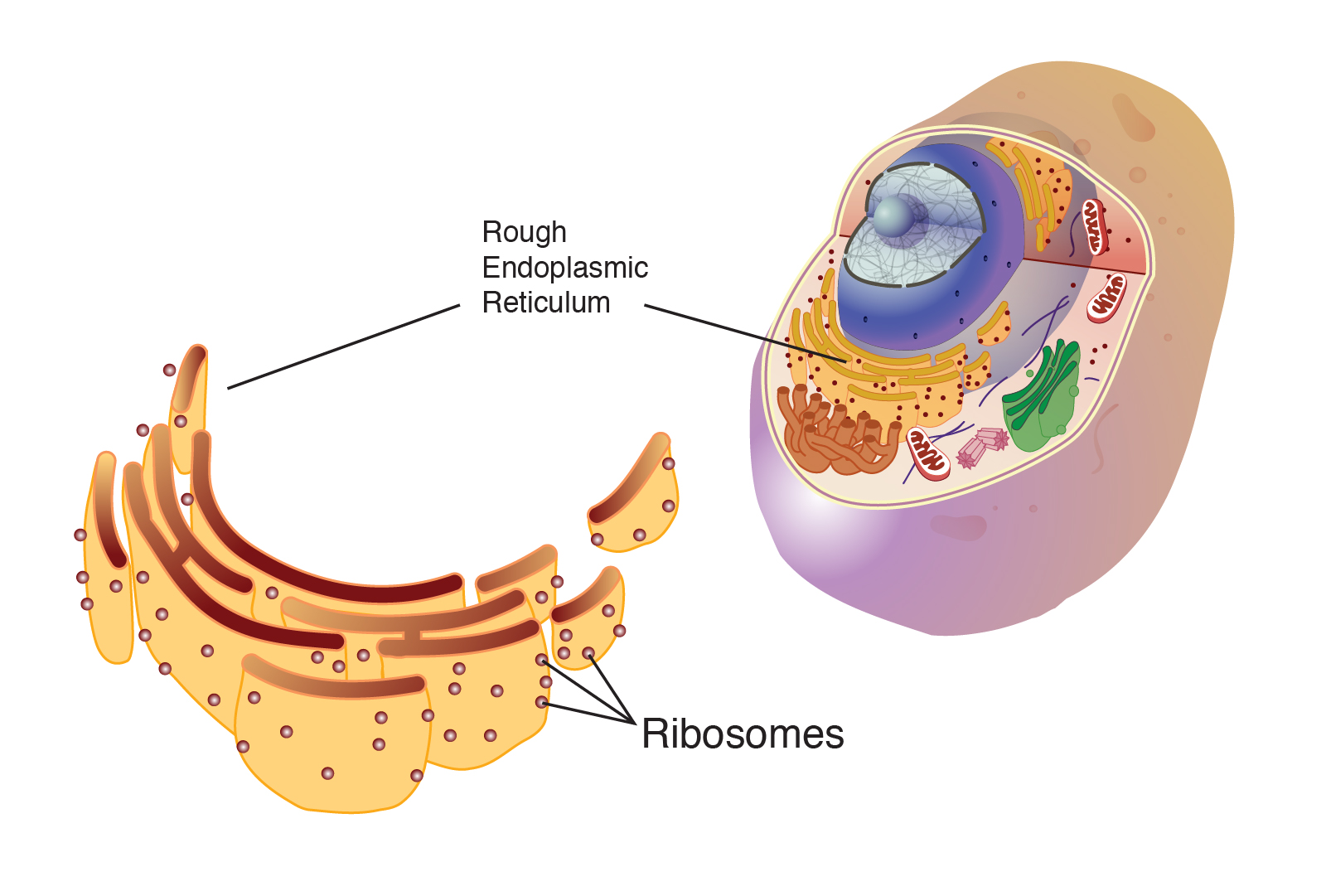
They are also found in the mitochondria and chloroplast of a. The mrna codes for specific amino acids and the ribosome is able to join the amino acids together in the specified order. For simplicity in this image, only the functional groups involved in the peptide bond are shown. Web figure 3.4.1 3.4. Ribosomes join amino acids together in a chain to form a protein ( figure 1 ). The word “synthesis” means “to combine things to produce something else.”. Web in this video i'm going to draw diagram of the ribosome, please send your feedback, #ribosome #ribosomediagram#sciencediagram 1 is an electron micrograph showing clusters of ribosomes. In prokaryotes, they float freely in the cytoplasm, while in eukaryotes they are free or remain bound to the outer membrane of the endoplasmic reticulum. Ribosomes are composed of special proteins and nucleic acids.
As catalysts they speed the time of reactions, as fibers they provide support, and many proteins function in specific tasks, like contracting muscle cells. Web a bacterial ribosome is about 250 nm in diameter and consists of two subunits, one large and one small. Web figure 3.4.1 3.4. Web the shape of a protein is what gives the protein its specific function. Amino acids are the subunits that are joined together by the ribosome to form a protein. The word “synthesis” means “to combine things to produce something else.”. The r and r' designations refer to the rest of each amino acid structure. The endoplasmic reticulum and golgi body are involved in protein maturation and transport. For simplicity in this image, only the functional groups involved in the peptide bond are shown. In prokaryotes, they float freely in the cytoplasm, while in eukaryotes they are free or remain bound to the outer membrane of the endoplasmic reticulum.
How to draw ribosomes YouTube
A single pancreas cell can synthesize 5 million molecules. Web figure 3.4.1 3.4. The r and r' designations refer to the rest of each amino acid structure. Web this video about ribosome drawing. Web ribosomes are the cellular structures responsible for protein synthesis.
Ribosome protein factory definition, function, structure and biology
The r and r' designations refer to the rest of each amino acid structure. A single pancreas cell can synthesize 5 million molecules. As catalysts they speed the time of reactions, as fibers they provide support, and many proteins function in specific tasks, like contracting muscle cells. In eukaryotes, the nucleolus is. Web a bacterial ribosome is about 250 nm.
Ribosomes Definition, Structure, & Functions, with Diagram
The r and r' designations refer to the rest of each amino acid structure. Ribosomes read mrna blueprints and piece together proteins, with the help of transfer rna (trna) support workers that bring them the right amino acid each step of the way. They are also found in the mitochondria and chloroplast of a. Web in this video i'm going.

RIBOSOME DRAWING BIOLOGY 9th class. YouTube
Web in this video i'm going to draw diagram of the ribosome, please send your feedback, #ribosome #ribosomediagram#sciencediagram Web ribosomes are cell structures present in large numbers in all living cells acting as the site of protein synthesis. Coli, there are between 10,000 and 70,000 ribosomes present in each cell at any given time.a ribosome is a complex macromolecule composed.

How to draw Ribosomes.. YouTube
These clusters, called polysomes, are held together by messenger rna (mrna). The endoplasmic reticulum and golgi body are involved in protein maturation and transport. A single pancreas cell can synthesize 5 million molecules. Web the shape of a protein is what gives the protein its specific function. The r and r' designations refer to the rest of each amino acid.

how to draw ribosome diagram easily YouTube
A peptide bond links the carboxyl end of one amino acid with the amino end of another, expelling one water molecule. Web this tutorial demonstrates how to draw ribosomes in powerpoint for research publication, conference posters, science figures and graphical abstracts. A peptide bond links the carboxyl end of one amino acid with the amino end of another, expelling one.
Ribosomes
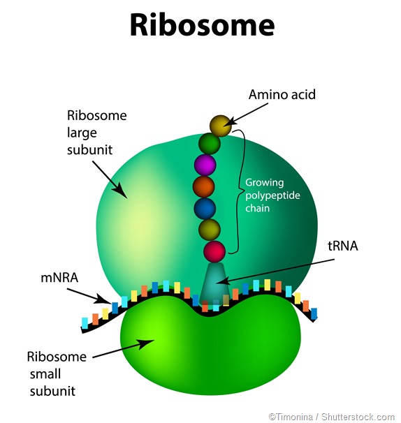
As the ribosome collects a new amino acid, it attaches this amino acid to the chain of amino acids it has just pieced together by performing a chemical reaction. The large subunit sits on top of the small subunit, with an rna template sandwiched between the two. A peptide bond links the carboxyl end of one amino acid with the.
Ribosome Structure
Web a ribosome is an organelle made of protein and rna in which protein synthesis occurs. Proteins are used in almost all cellular functions; The structure of prokaryotic ribosome is given in the figure 8.2 b. Web this reaction is catalyzed by ribosomes and generates one water molecule. The mrna codes for specific amino acids and the ribosome is able.
Ribosome
Sure enough, they found that ribosomes that build the pol protein stopped just before the very end, leaving the protein tethered to the ribosome like a balloon tied to a child’s hand. Web a ribosome is an organelle made of protein and rna in which protein synthesis occurs. Proteins are used in almost all cellular functions; As the ribosome collects.
.jpg)
AS Biology The appearance, ultrastructure and function of ribosomes.
Web in this video i'm going to draw diagram of the ribosome, please send your feedback, #ribosome #ribosomediagram#sciencediagram Ribosomes read mrna blueprints and piece together proteins, with the help of transfer rna (trna) support workers that bring them the right amino acid each step of the way. Web this video about ribosome drawing. They can make up 25% of the.
The Mrna Codes For Specific Amino Acids And The Ribosome Is Able To Join The Amino Acids Together In The Specified Order.
For simplicity in this image, only the functional groups involved in the peptide bond are shown. In splicing, some sections of the rna transcript ( introns) are removed, and the remaining. The translation of information and the linking of amino acids are at the heart of the protein production process.a ribosome, formed from two subunits locking together, functions to: Web multiple prolines in a row can stop a ribosome in its tracks.
The R And R' Designations Refer To The Rest Of Each Amino Acid Structure.
Web ribosomes are cell structures present in large numbers in all living cells acting as the site of protein synthesis. They can make up 25% of the dry weight of cells (e.g., pancreas cells) and specialize in protein synthesis. Coli, there are between 10,000 and 70,000 ribosomes present in each cell at any given time.a ribosome is a complex macromolecule composed of structural and catalytic rrnas, and many distinct polypeptides. Web a bacterial ribosome is about 250 nm in diameter and consists of two subunits, one large and one small.
How To Draw Golgi Body And Ribosomes | How To Draw Diagram Of Golgi Body With Ribosomeshello Friends In This Video I Tell You About How To Draw Golgi.
The endoplasmic reticulum and golgi body are involved in protein maturation and transport. All proteins start as deoxyribonucleic acid, or dna. As catalysts they speed the time of reactions, as fibers they provide support, and many proteins function in specific tasks, like contracting muscle cells. The colored balls at the top of this diagram represent different amino acids.
Vacuoles Are Storage Compartments That Sequester Waste And Help Maintain Water.
They are also found in the mitochondria and chloroplast of a. Even before an mrna is translated, a cell must invest energy to build each of its ribosomes. Web in this video i'm going to draw diagram of the ribosome, please send your feedback, #ribosome #ribosomediagram#sciencediagram The structure of prokaryotic ribosome is given in the figure 8.2 b.