Human Heart Drawing And Label
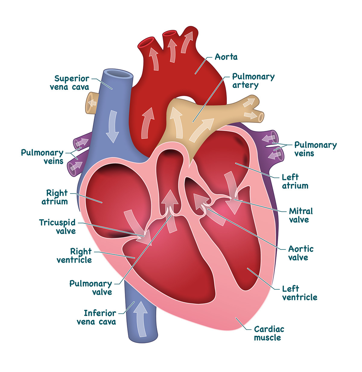
Human Heart Drawing And Label - Each of the upper chambers, the right atrium (plural = atria) and the left atrium, acts as a receiving chamber and contracts to push blood into the lower chambers, the right ventricle and the left ventricle. New 3d rotate and zoom. Selecting or hovering over a box will highlight each area in the diagram. Web to draw the internal structure of the heart, start by sketching the 2 pulmonary veins to the lower left of the aorta and the bottom of the inferior vena cava slightly to the right of that. Images are labelled, providing an invaluable medical and anatomical tool. It pumps blood from the heart to different parts of the body and back to the heart. Web in this lecture, dr mike shows the two best ways to draw and label the heart! This is a file from the wikimedia commons. Controls the rhythm and speed of your heart rate. The human heart and its functions are truly fascinating.
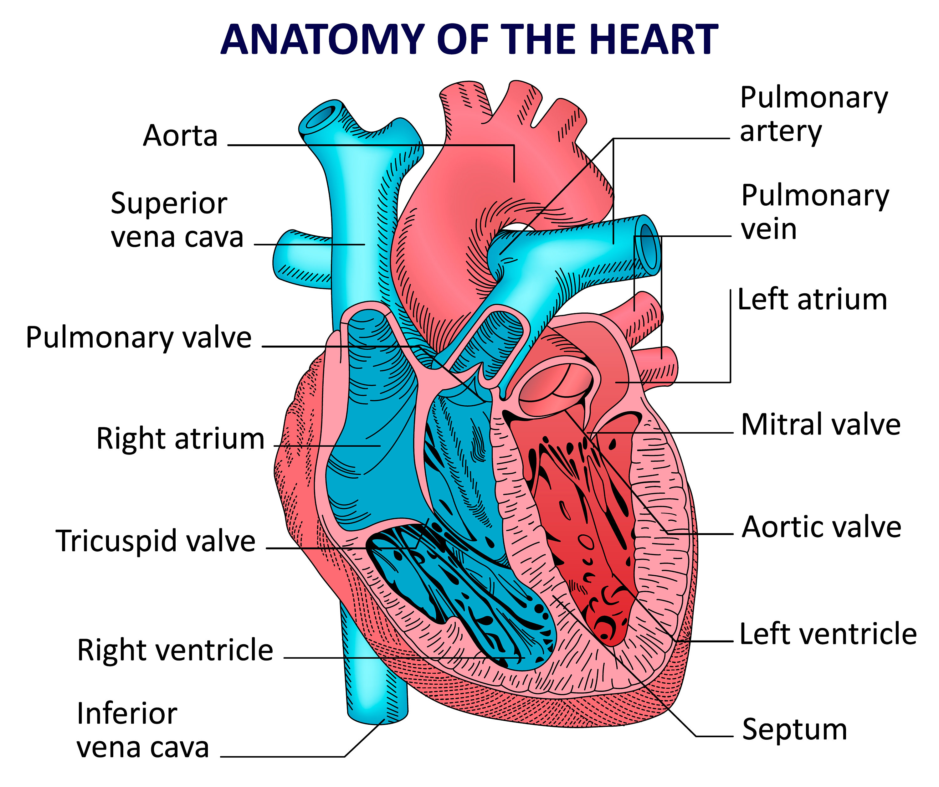
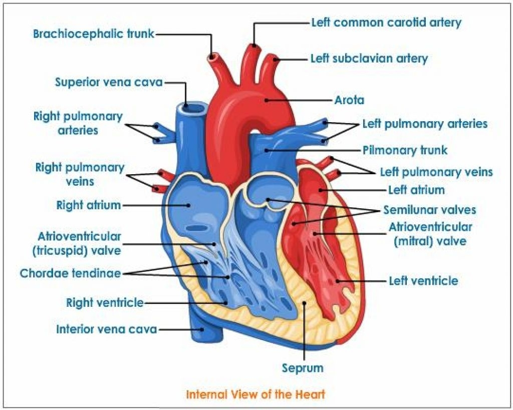
The heart is a mostly hollow, muscular organ composed of cardiac muscles and. After all, we know that stress is bad for the heart! This tool provides access to several medical illustrations, allowing the user to interactively discover heart anatomy. Web welcome to the anatomy of the heart made easy! Original file (svg file, nominally 663 × 651 pixels, file size: It's situated a little to the left of your chest center, and it's around your fist size. For this first step of our guide on how to draw a human heart, we will start with some outlines for the heart. New 3d rotate and zoom. Drag and drop the text labels onto the boxes next to the diagram. Cross section of the human heart.
It's situated a little to the left of your chest center, and it's around your fist size. Web to draw the internal structure of the heart, start by sketching the 2 pulmonary veins to the lower left of the aorta and the bottom of the inferior vena cava slightly to the right of that. In this interactive, you can label parts of the human heart. Web the human heart is the most crucial organ of the human body. Anatomy of the heart [10:27] overview of the anatomy and functions of the heart. Web the human heart. Moreover, the heart lies under the rib cage, in the left of the breastbone (sternum) and the right behind the lungs and above the diaphragm. Heart (right lateral view) the heart is a muscular organ that pumps blood around the body by circulating it through the circulatory/vascular system. New 3d rotate and zoom. In addition to reviewing the human heart anatomy, we will also discuss the function and order in which blood flows through the heart.
Heart And Labels Drawing at GetDrawings Free download
It also takes away carbon dioxide and other waste so other organs can dispose of them. 1.1m views 3 years ago drawing tutorials. It pumps blood from the heart to different parts of the body and back to the heart. This tool provides access to several medical illustrations, allowing the user to interactively discover heart anatomy. Web drawing a human.
human heart drawing labeled
Web anatomy of the human heart and coronaries: In this interactive, you can label parts of the human heart. For this first step of our guide on how to draw a human heart, we will start with some outlines for the heart. Web drawing a human heart is easier than you may think. 14 views 1 year ago.
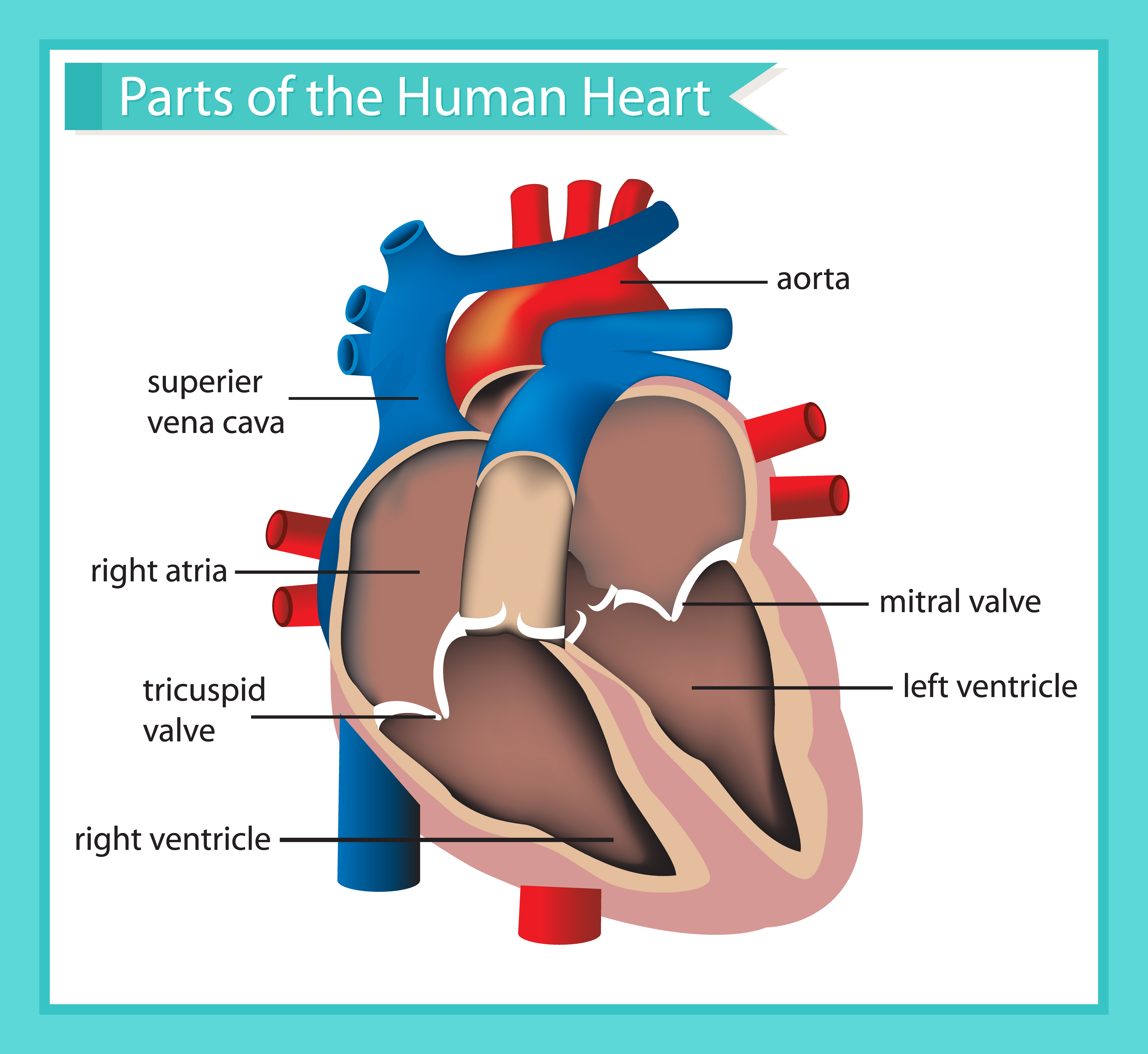
Scientific medical illustration of parts of the human heart 685453
Plus, you may just learn something new along the way. Original file (svg file, nominally 663 × 651 pixels, file size: 93 kb) render this image in. It's situated a little to the left of your chest center, and it's around your fist size. Anatomy of the heart [10:27] overview of the anatomy and functions of the heart.
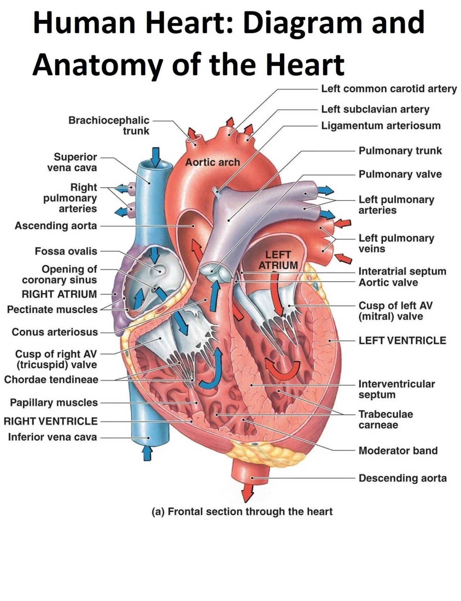
The Human Heart Diagram Display Poster Diagram and Anatomy of the Heart
In this interactive, you can label parts of the human heart. The heart is a muscle. Web drawing a human heart is easier than you may think. New 3d rotate and zoom. Cross section of the human heart.

31 Human Heart To Label Labels Design Ideas 2020
Then, fill in the base of the heart with the. Do you want a fun way to learn the structure of the heart? Blood brings oxygen and nutrients to your cells. Heart (right lateral view) the heart is a muscular organ that pumps blood around the body by circulating it through the circulatory/vascular system. Original file (svg file, nominally.

heart anatomy labeling
Heart (right lateral view) the heart is a muscular organ that pumps blood around the body by circulating it through the circulatory/vascular system. By following the simple steps, you too can easily draw a perfect human heart. Each of the upper chambers, the right atrium (plural = atria) and the left atrium, acts as a receiving chamber and contracts to.
Heart And Labels Drawing at GetDrawings Free download
So, grab your pencil and sketchbook because we’ve got a heart to draw! Web cardiovascular heart diagram: Original file (svg file, nominally 663 × 651 pixels, file size: Use some curved lines for this aorta with a. Moreover, the heart lies under the rib cage, in the left of the breastbone (sternum) and the right behind the lungs and.

humanheartdiagram Tim's Printables
Do you want a fun way to learn the structure of the heart? It pumps blood from the heart to different parts of the body and back to the heart. In this interactive, you can label parts of the human heart. How to visualize anatomic structures. We will use labeled diagrams and pictures to learn the main cardiac structures and.
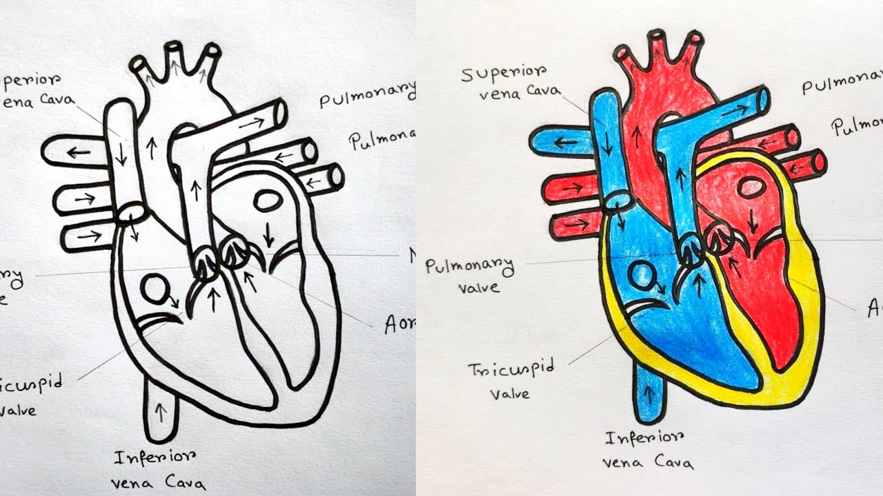
How to draw Human Heart with colour Human Heart labelled diagram
Web drawing a human heart is easier than you may think. 336k views 1 year ago easy diagrams drawings. Web medically reviewed by the healthline medical network — by the healthline editorial team — updated on january 20, 2018. The most common heart attack symptoms or warning signs are chest pain, breathlessness, nausea, sweating etc. The heart is a muscle.

How To Draw Human Heart Diagram
This is a file from the wikimedia commons. Cross section of the human heart. 336k views 1 year ago easy diagrams drawings. The most common heart attack symptoms or warning signs are chest pain, breathlessness, nausea, sweating etc. By following the simple steps, you too can easily draw a perfect human heart.
Each Of The Upper Chambers, The Right Atrium (Plural = Atria) And The Left Atrium, Acts As A Receiving Chamber And Contracts To Push Blood Into The Lower Chambers, The Right Ventricle And The Left Ventricle.
93 kb) render this image in. 336k views 1 year ago easy diagrams drawings. This is a file from the wikimedia commons. It also takes away carbon dioxide and other waste so other organs can dispose of them.
How To Visualize Anatomic Structures.
For this first step of our guide on how to draw a human heart, we will start with some outlines for the heart. Get free printable coloring page of this drawing. The heart is a mostly hollow, muscular organ composed of cardiac muscles and. The left side and the right side each have one atrium and one ventricle.
The Heart, One Of The Most Significant Organs In The Human Body, Is Nothing But A Muscular Pump Which Pumps Blood Throughout The Body.
Web in this lecture, dr mike shows the two best ways to draw and label the heart! Size of this png preview of this svg file: This tool provides access to several medical illustrations, allowing the user to interactively discover heart anatomy. Web the human heart consists of four chambers:
Use Some Curved Lines For This Aorta With A.
The most common heart attack symptoms or warning signs are chest pain, breathlessness, nausea, sweating etc. Web welcome to the anatomy of the heart made easy! Selecting or hovering over a box will highlight each area in the diagram. Images are labelled, providing an invaluable medical and anatomical tool.