Hyaline Cartilage Drawing
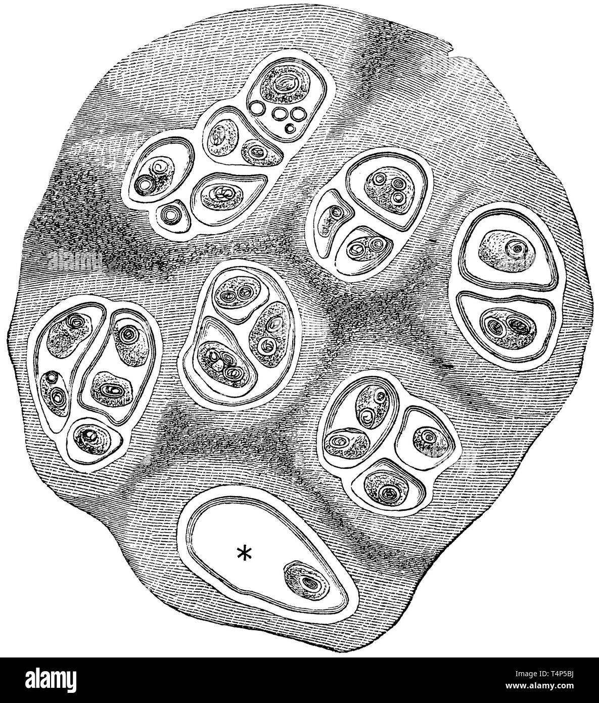
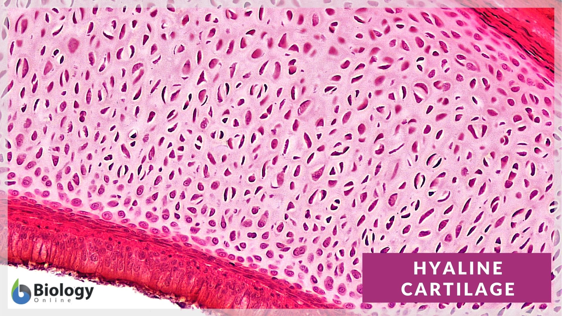
Hyaline Cartilage Drawing - Isogenous groups and interstitial growth results when chondrocytes divide and produce extracellular matrix. Web during embryonic development, hyaline cartilage serves as temporary cartilage models that are essential precursors to the formation of most of the axial and appendicular skeleton. Each slide is shown with additional information to its right. This post will describe the basic histology of hyaline cartilage with slide images and labeled diagram. This image shows a cross section of a cartilage ring that supports the trachea and maintains the. The image can be changed using any combination of the following. Medical school university of minnesota minneapolis, mn. The word hyaline is derived from the greek word ‘ hyalos ’, which means ‘ glassy ’ implying its shiny, smooth appearance. Medical school university of minnesota minneapolis, mn. A joint of the jaw that connects it to the temporal bones of the skull.
Web the hyaline cartilage in the trachea is in the middle of the tracheal wall. Hyaline cartilage is the most prevalent type, forming articular cartilages and the framework for parts of the nose, larynx, and trachea. The image can be changed using any combination of the following commands. The lack of blood vessels in hyaline cartilage means that nutrients and wastes must diffuse through the tissue, thus limiting the thickness of the hyaline cartilage. Use the image slider below to learn how to use a microscope to identify and study hyaline cartilage on a microscope slide of the trachea. It is the most common cartilage and is found on articular surfaces of bone, walls of the respiratory system (trachea and bronchi), and epiphyseal plates. Step by step drawing of histology of hyaline cartilage A joint of the jaw that connects it to the temporal bones of the skull. It tends to stain more blue than other kinds of connective tissue (however, remember that color should never be the main cue you use to identify a tissue). Web likecomment share subscribe #hyalinecartilage #histodiagrams #hyalinecartilagediagram #cartilagehistology
This post will describe the basic histology of hyaline cartilage with slide images and labeled diagram. Star star star star star. Web likecomment share subscribe #hyalinecartilage #histodiagrams #hyalinecartilagediagram #cartilagehistology Hyaline cartilage is the most prevalent type, forming articular cartilages and the framework for parts of the nose, larynx, and trachea. You can begin to see the details in hyaline cartilage (hc. A type of cartilage found on many joint surfaces; This section comes from cartilage in a developing fetal bone. Web 48,960 x 39,529 pixels 7.2 gb. A histological overview of the most common type of cartilage in the human body. Medical school university of minnesota minneapolis, mn.
Hyaline cartilage hires stock photography and images Alamy
A histological overview of the most common type of cartilage in the human body. Medical school university of minnesota minneapolis, mn. Star star star star star. Hyaline cartilage is the most prevalent type, forming articular cartilages and the framework for parts of the nose, larynx, and trachea. Each slide is shown with additional information to its right.
Hyaline Cartilage Cells ClipArt ETC
Isogenous groups and interstitial growth results when chondrocytes divide and produce extracellular matrix. This is a section of hyaline cartilage, the most abundant type of cartilage in the body. This image shows a cross section of a cartilage ring that supports the trachea and maintains the. Each slide is shown with additional information to its right. Click on links to.

Illustrations Hyaline Cartilage General Histology
A histological overview of the most common type of cartilage in the human body. Web the hyaline cartilage in the trachea is in the middle of the tracheal wall. A type of cartilage found on many joint surfaces; It tends to stain more blue than other kinds of connective tissue (however, remember that color should never be the main cue.
Long Bone Diagram Hyaline Cartilage / Anatomy and Phisology PP
This rapidly growing tissue forms the majority of the fetal skeleton and remains into adulthood as the smooth joint surfaces at the. Web during embryonic development, hyaline cartilage serves as temporary cartilage models that are essential precursors to the formation of most of the axial and appendicular skeleton. Use the hotspot image below to learn more about the characteristics of.
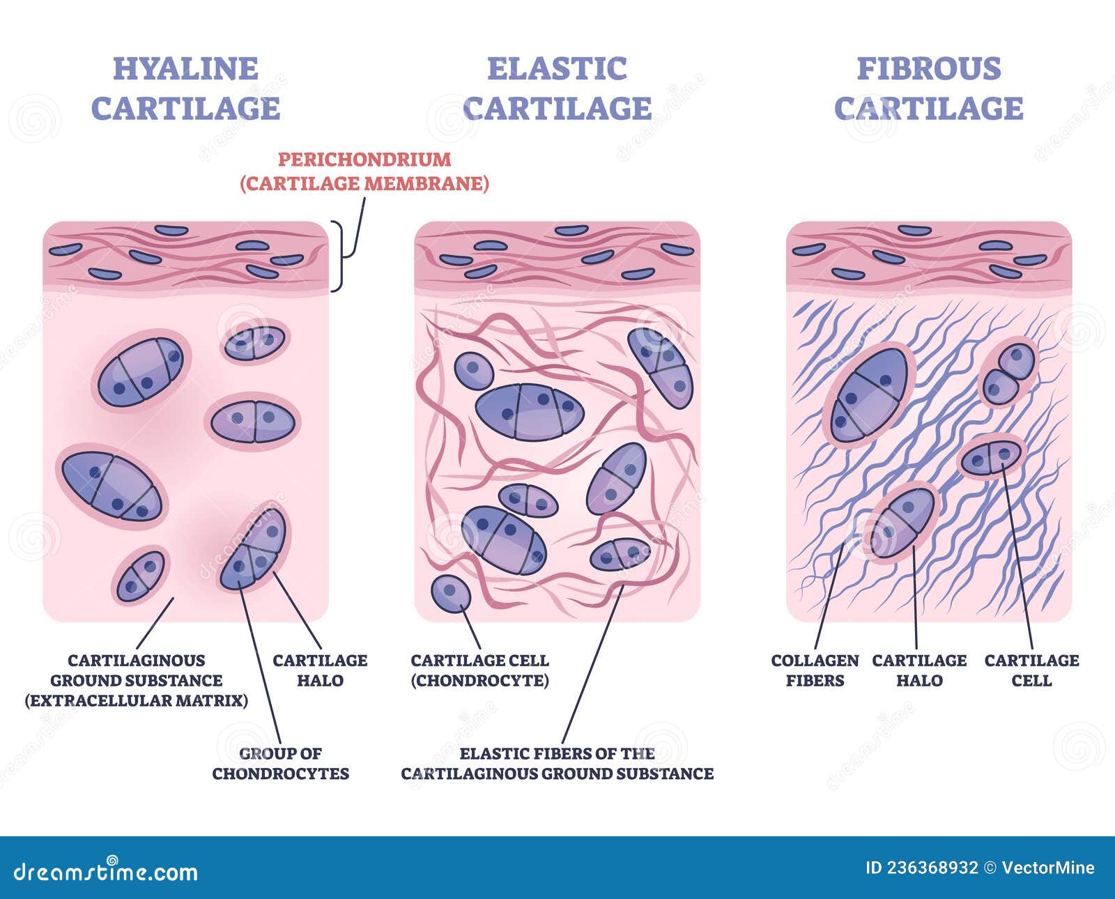
Perichondrium As Hyaline and Elastic Cartilage Membrane Outline Diagram
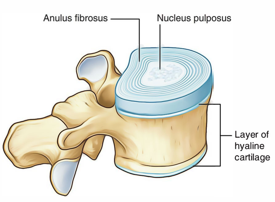
The bar shows the position of the hyaline cartilage. This rapidly growing tissue forms the majority of the fetal skeleton and remains into adulthood as the smooth joint surfaces at the. It contains no nerves or blood vessels, and its structure is relatively simple. Each slide is shown with additional information to its right. The image can be changed using.

Hyaline Cartilage Labeled Diagram
Step by step drawing of histology of hyaline cartilage Mh 046 hyaline articular cartilage. A type of cartilage found on many joint surfaces; This rapidly growing tissue forms the majority of the fetal skeleton and remains into adulthood as the smooth joint surfaces at the. Medical school university of minnesota minneapolis, mn.

Schematic drawing of articular (hyaline) cartilage containing abundant
You can begin to see the details in hyaline cartilage (hc. Use the image slider below to learn how to use a microscope to identify and study hyaline cartilage on a microscope slide of the trachea. Web during embryonic development, hyaline cartilage serves as temporary cartilage models that are essential precursors to the formation of most of the axial and.
Connective Tissue Supports and Protects · Anatomy and Physiology
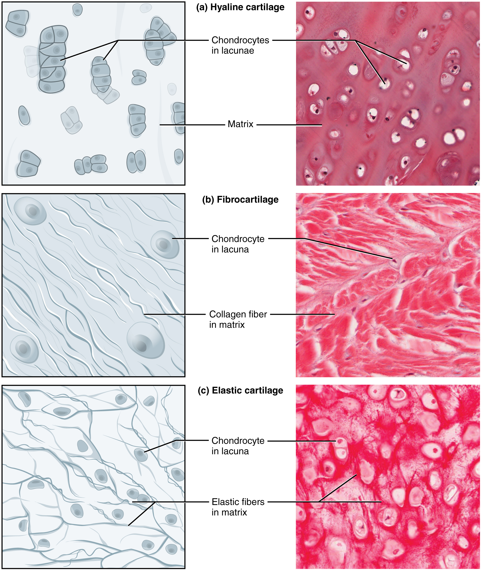
Cells that form and maintain the cartilage. A type of cartilage found on many joint surfaces; Star star star star star. The word hyaline is derived from the greek word ‘ hyalos ’, which means ‘ glassy ’ implying its shiny, smooth appearance. Web the hyaline cartilage in the trachea is in the middle of the tracheal wall.

Hyaline Cartilage Drawing YouTube
It tends to stain more blue than other kinds of connective tissue (however, remember that color should never be the main cue you use to identify a tissue). This article will focus on important features of hyaline cartilage, namely its matrix, chondrocytes, and perichondrium. Cells that form and maintain the cartilage. This section comes from cartilage in a developing fetal.
Long Bone Diagram Hyaline Cartilage What Is Cartilage
It contains no nerves or blood vessels, and its structure is relatively simple. It is also most commonly found in the ribs, nose, larynx, and trachea. Web the hyaline cartilage in the trachea is in the middle of the tracheal wall. This article will focus on important features of hyaline cartilage, namely its matrix, chondrocytes, and perichondrium. Each slide is.
It Is Also Most Commonly Found In The Ribs, Nose, Larynx, And Trachea.
Hyaline cartilage is a type of connective tissue found in areas such as the nose, ears, and trachea of the human body. Cartilage is flexible connective tissue found throughout the whole body. A joint of the jaw that connects it to the temporal bones of the skull. Hyaline cartilage is the most prevalent type, forming articular cartilages and the framework for parts of the nose, larynx, and trachea.
The Image Can Be Changed Using Any Combination Of The Following Commands.
This rapidly growing tissue forms the majority of the fetal skeleton and remains into adulthood as the smooth joint surfaces at the. Web likecomment share subscribe #hyalinecartilage #histodiagrams #hyalinecartilagediagram #cartilagehistology It tends to stain more blue than other kinds of connective tissue (however, remember that color should never be the main cue you use to identify a tissue). Medical school university of minnesota minneapolis, mn.
It Contains No Nerves Or Blood Vessels, And Its Structure Is Relatively Simple.
This section comes from cartilage in a developing fetal bone. This image shows a cross section of a cartilage ring that supports the trachea and maintains the. Click on links to move to a. The image can be changed using any combination of the following.
Watch The Video Tutorial Now.
Mh 046 hyaline articular cartilage. Star star star star star. This article will focus on important features of hyaline cartilage, namely its matrix, chondrocytes, and perichondrium. Each slide is shown with additional information to its right.