Label The Schematic Drawing Of A Kidney
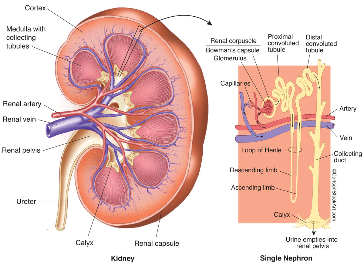
Label The Schematic Drawing Of A Kidney - Web simple labeling exercise on the structure of the kidney. Learn vocabulary, terms, and more with flashcards, games, and other study tools. Each kidney is approximately three vertebrae in length. Learn vocabulary, terms, and more with flashcards, games, and other study tools. Also, the diagram shows the relationship between the. Web anatomy of the urinary system. Surrounding the kidney tubules of cortical nephrons and the proximal and distal convoluted tubules of juxtamedullary nephrons There are about 1,000,000 nephrons in each human kidney. Web this online quiz is called label the kidney. Let us look at the components of the nephron.
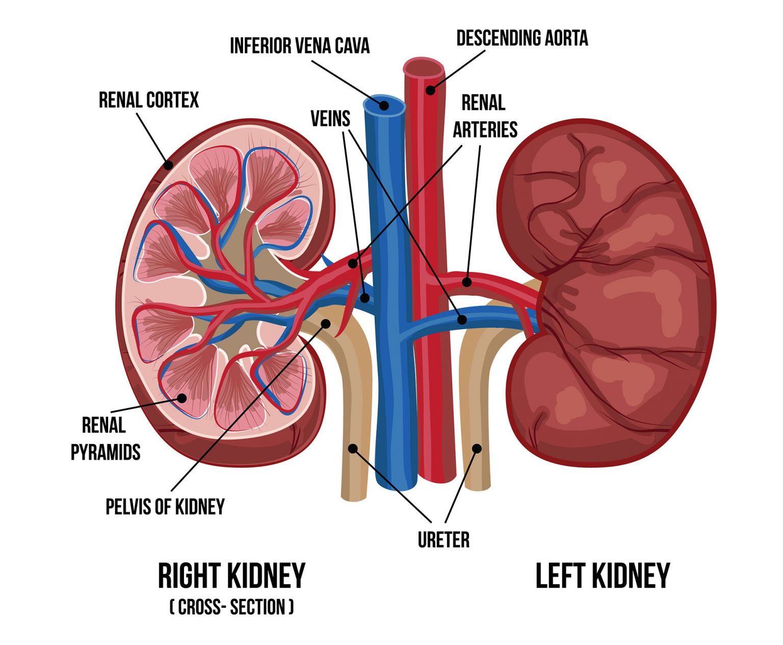
Front view diagram of the urinary tract. Drag the labels onto the diagram to identify the parts of the renal corpuscle. Ureter shown connecting with the pelvis / on concave side /. Web drag the labels to identify the organs of the urinary system. Learn vocabulary, terms, and more with flashcards, games, and other study tools. Labels point to the kidney, ureter, bladder, and urethra. The human kidneys house millions of tiny filtration units called nephrons, which enable our body to retain the vital nutrients, and excrete the unwanted or excess molecules as well as metabolic wastes from the body. The urinary system consists of the kidneys, ureters, urinary bladder, and urethra. Web microscopic structures mostly found in the renal cortex, but some components found in the medulla; The kidneys lie retroperitoneally (behind the peritoneum) in the abdomen, either side of the vertebral column.
Surrounding the kidney tubules of cortical nephrons and the proximal and distal convoluted tubules of juxtamedullary nephrons Web terms in this set (13) start studying kidney label drawing. There are about 1,000,000 nephrons in each human kidney. Students drag labels to the structures on the slide. This online quiz is called label the kidney. This area of the kidney is called the hilum. Web microscopic structures mostly found in the renal cortex, but some components found in the medulla; Web terms in this set (11) a duct leading from the kidney to the urinary bladder. The video also highlights the role of the renal cortex and renal medulla in. Ureter shown connecting with the pelvis / on concave side /.
Human kidney anatomy diagram 446409 Vector Art at Vecteezy
Web anatomy of the urinary system. The kidneys filter the blood to remove wastes and produce urine. Learn vocabulary, terms, and more with flashcards, games, and other study tools. By the end of this section, you will be able to: Pelvis shown on the concave side of the kidney;
Label the Parts of the Urinary System
Each kidney is approximately three vertebrae in length. Drag the labels onto the diagram to identify the parts of the renal corpuscle. It was created by member mpurzycki and has 9 questions. Web simple labeling exercise on the structure of the kidney. Let us look at the components of the nephron.

Human kidney anatomy diagram Royalty Free Vector Image
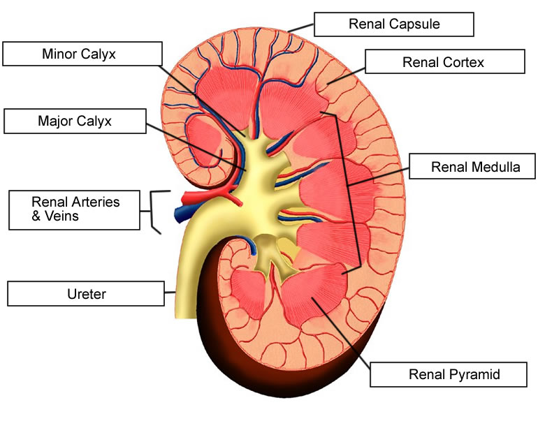
Web 11.3.2 draw and label a diagram of the kidney. Filters the blood to remove wastes and fluid; Web this article covers the anatomy of the kidneys, their function and internal structure together with the nephron. It was created by member mpurzycki and has 9 questions. Use listed terms (ureter, calyx, vessels.) to label each area of the kidney and.
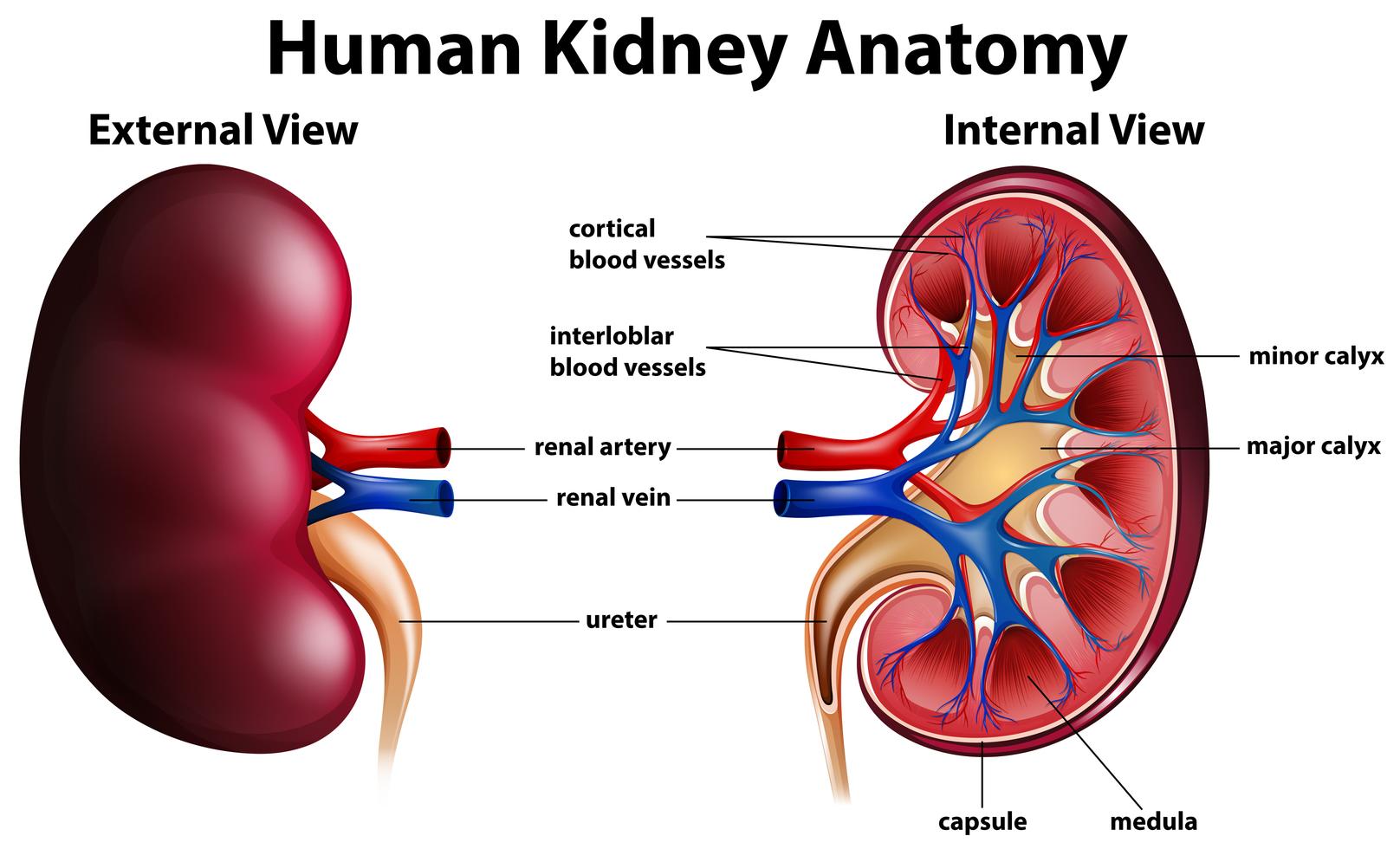
Diagram showing human kidney anatomy 295196 Vector Art at Vecteezy
Drag the labels onto the diagram to identify the blood vessels of the kidneys. Learn vocabulary, terms, and more with flashcards, games, and other study tools. Web terms in this set (13) start studying kidney label drawing. The urinary system consists of the kidneys, ureters, urinary bladder, and urethra. Web this online quiz is called label the kidney.
5b2 Organs HumanBio
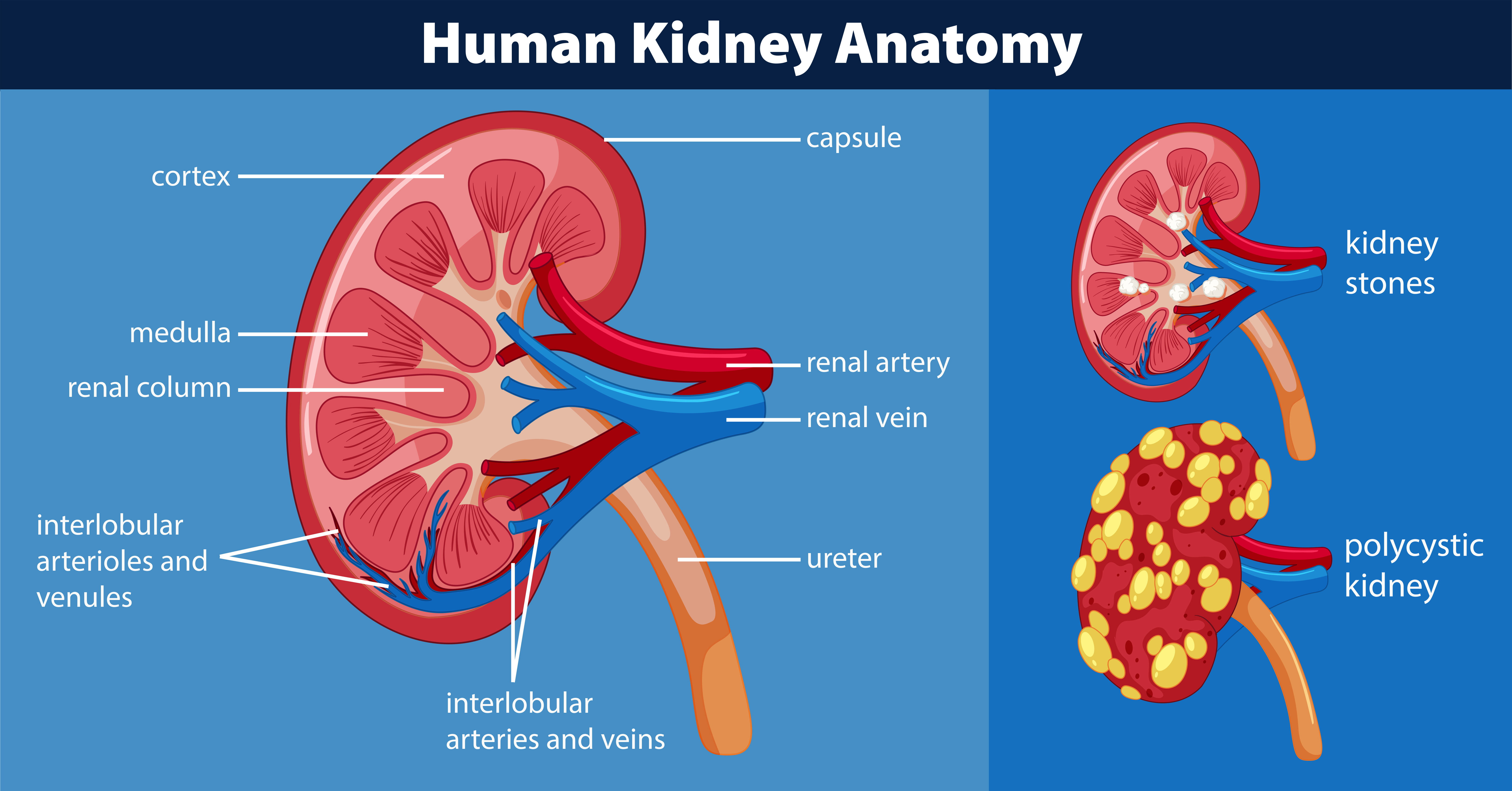
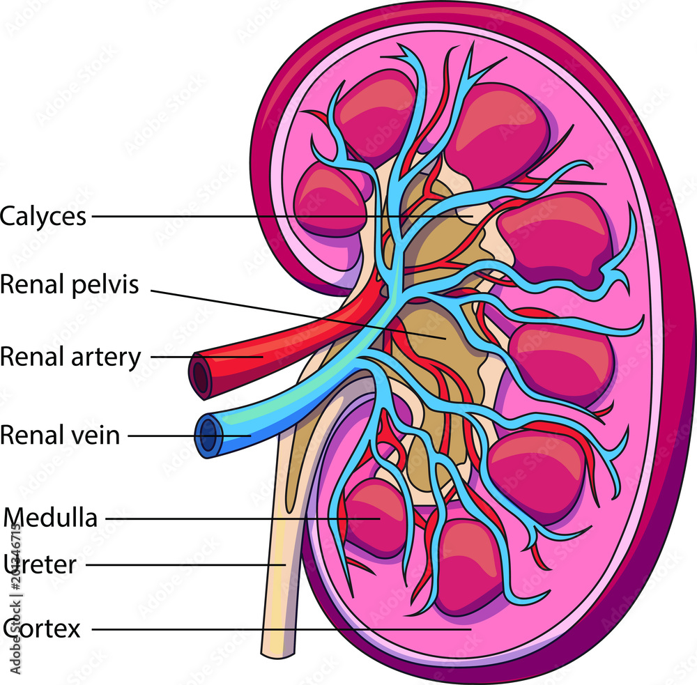
Ureter shown connecting with the pelvis / on concave side /. You can see this clearly in the detailed diagram of kidney anatomy shown in figure \(\pageindex{3}\). This problem has been solved! New 3d rotate and zoom. Cortex shown at the edge of kidney;

Human kidney medical diagram with a cross section Vector Image
They are roughly the size of your fist, and the male kidney. The kidneys lie retroperitoneally (behind the peritoneum) in the abdomen, either side of the vertebral column. This problem has been solved! Surrounding the kidney tubules of cortical nephrons and the proximal and distal convoluted tubules of juxtamedullary nephrons The concave side is where the renal artery enters the.

Labeled Diagram of the Human Kidney
Nephron is a microscopic structure that is the functional unit of the kidney. Learn vocabulary, terms, and more with flashcards, games, and other study tools. They are roughly the size of your fist, and the male kidney. This online quiz is called label the kidney. Web start studying kidney anatomy labeling.
Anatomy Of Kidney Nephron Anatomical Charts & Posters
Identify the major internal divisions and structures of the kidney. The video also highlights the role of the renal cortex and renal medulla in. This problem has been solved! Label the schematic drawing of a kidney. Students drag labels to the structures on the slide.

Diagram of human kidney anatomy Royalty Free Vector Image
New 3d rotate and zoom. Also, the diagram shows the relationship between the. Describe the macroscopic and microscopic anatomy of the kidney. They are roughly the size of your fist, and the male kidney. Web terms in this set (11) a duct leading from the kidney to the urinary bladder.
Schematic vector diagram of a kidney. Kidney structure with labeled
Web the video provides a detailed overview of the kidney's smallest functional unit, the nephron. By the end of this section, you will be able to: Medulla shown inside the cortex (with pyramids); Also, the diagram shows the relationship between the. Each kidney is approximately three vertebrae in length.
Web Drag The Labels To Identify The Organs Of The Urinary System.
The video also highlights the role of the renal cortex and renal medulla in. Medulla shown inside the cortex (with pyramids); Each kidney is approximately three vertebrae in length. This online quiz is called label the kidney.
The Urinary System Consists Of The Kidneys, Ureters, Urinary Bladder, And Urethra.
Ureter shown connecting with the pelvis / on concave side /. The kidneys filter the blood to remove wastes and produce urine. Web this article covers the anatomy of the kidneys, their function and internal structure together with the nephron. Let us look at the components of the nephron.
Learn Vocabulary, Terms, And More With Flashcards, Games, And Other Study Tools.
Free kidney labeling quiz for students biology, anatomy and physiology. They typically extend from t12 to l3, although the right kidney is often situated slightly lower due to the presence of the liver. The human kidneys house millions of tiny filtration units called nephrons, which enable our body to retain the vital nutrients, and excrete the unwanted or excess molecules as well as metabolic wastes from the body. Start studying correctly label the following anatomical parts of a kidney.
Pelvis Shown On The Concave Side Of The Kidney;
You'll get a detailed solution from a subject matter expert that helps you learn core concepts. Identify the major internal divisions and structures of the kidney. Urethra minor calyx renal pelvis renal medulla renal pyramid ureter renal cortex major calyx. Learn vocabulary, terms, and more with flashcards, games, and other study tools.