Medulla Drawing
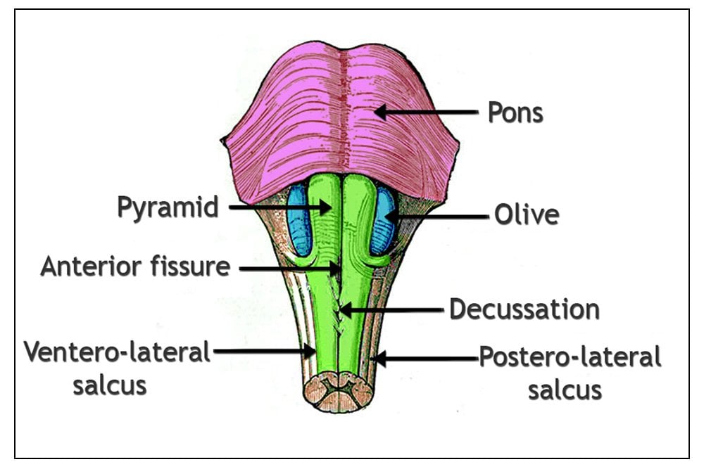
Medulla Drawing - • specify its medullary pyramids, which contain descending motor fibers. Their shape resembles a bean, where we can describe the superior and inferior poles, as well as the major convexity pointed laterally, and the minor concavity pointed. Web draw a pair of rectangles in the center. Web the medulla oblongata plays a critical role in transmitting signals between the spinal cord and the higher parts of the brain and in controlling autonomic activities, such as heartbeat and respiration. Schematic drawing of the pig brain, indicating the location of medulla oblongata from a sagittal view. Web medulla oblongata is situated inferior to the pons. Describe the morphology of the juxtaglomerular apparatus and explain its function during regulation of the circulatory and renal systems. Cross section at the decussation of the pyramids. Place the circle on the bottom line of the oval and draw a line connecting the circle to the oval outline. Motor neurons cross from the left.
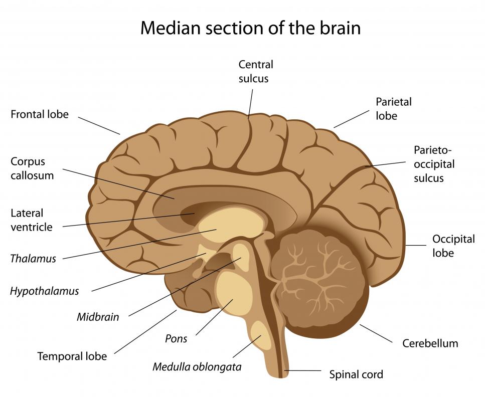
This neuroanatomical atlas is therefore perfectly adapted for the web. And then the hind brain will become the rest of the brain. Medial view of the left hemisphere of the human brain. Web it is composed of 64 drawings, illustrations and anatomical charts, all in vector format. Web that finishes this session. Try to draw the medulla and mark medial and lateral medullary syndromes. It is situated inside the vertebral canal of the vertebral column. • denote that, from a clinician's broad perspective, the medulla is the center for the most basic generators of life. Web medulla oblongata is situated inferior to the pons. The kidneys are bilateral organs placed retroperitoneally in the upper left and right abdominal quadrants and are part of the urinary system.
Web the medulla oblongata plays a critical role in transmitting signals between the spinal cord and the higher parts of the brain and in controlling autonomic activities, such as heartbeat and respiration. Next time you see a patient, try to draw the lesion before seeing the imaging. The brain stem was represented on multiple angles, to show the external structures of the midbrain, pons and medulla oblongata (bulb) and a projection of the cranial nerve nuclei. Describe the structure of the renal cortex vs. Medulla and correlate it with functions of both. • first, draw the different brainstem levels, from superior to inferior: Web the medulla oblongata. It is a conduit for many ascending and descending nerve tracts that carry the information between the brain and spinal cord. Web the medulla overview here, we'll learn about the medulla. Web the midbrainoverview here, we'll learn about the midbrain.
:max_bytes(150000):strip_icc()/GettyImages-1092334754-a7bfe7bc880e433d86d9354095012158.jpg)
Die Anatomie der Medulla Oblongata MedDe
First, draw the different brainstem levels, from superior to inferior: Motor neurons cross from the left. Web how to draw the level of decussation of pyramids in medulla oblongata. It is situated inside the vertebral canal of the vertebral column. The medulla oblongata (medulla) is one of the three regions that make up the brainstem.

What Are the Medullary Pyramids? (with pictures)
And then the hind brain will become the rest of the brain. Cross section at the decussation of the pyramids. Medulla and correlate it with functions of both. Web draw a pair of rectangles in the center. Web the medulla overview here, we'll learn about the medulla.

The Medulla Oblongata Internal Structure Vasculature TeachMeAnatomy
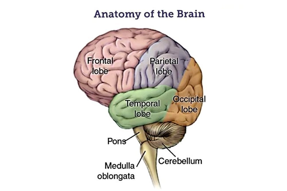
• midbrain • pons • medulla label the anterior/posterior orientational plane. Try to draw the medulla and mark medial and lateral medullary syndromes. Web the medulla oblongata has many important features and functions. The pons, the medulla, and the cerebellum. During development, there’s a disproportion between spinal cord growth and vertebral column growth.
Medulla Oblongata Definition, Structure And Functions
They're from the developing nervous system. Cross section at the decussation of the pyramids. During development, there’s a disproportion between spinal cord growth and vertebral column growth. Web the midbrainoverview here, we'll learn about the midbrain. Medulla and correlate it with functions of both.

Vertical section of a human brain. showing the medulla, pons
If it's easier for you, draw a horizontal oval and make a circle that's about 1/3 of the size of the bottom line. Medulla and correlate it with functions of both. First, draw the different brainstem levels, from superior to inferior: The easy and simple drawing of pyram. Sketch a narrow curve above the top line of the brain.
What Are the Different Parts of the Medulla Oblongata?
In the brainstem, one usually get excellent clinical imaging correlation, and if you have examined properly, your localisation will be correct most of the time. The renal medulla contains the following structures: The brain stem was represented on multiple angles, to show the external structures of the midbrain, pons and medulla oblongata (bulb) and a projection of the cranial nerve.
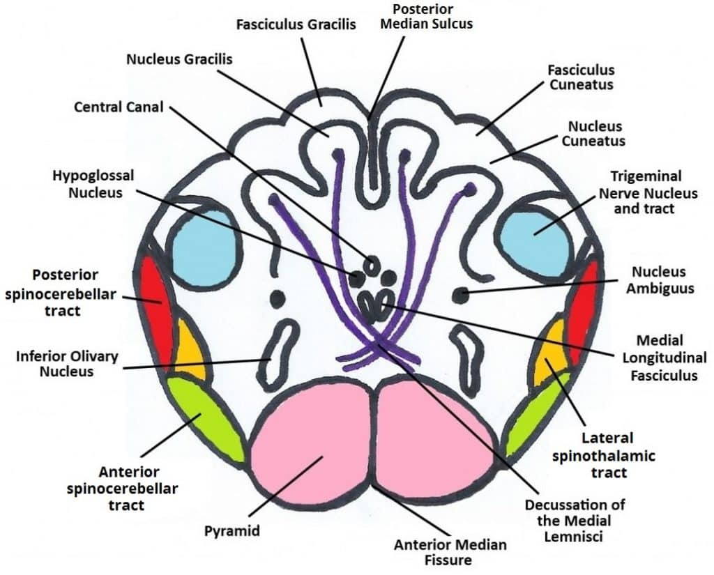
Image Gallery medulla anatomy
Describe the structure of each segment of the nephron. Web • to begin, draw the cervical spinal cord. • first, draw the different brainstem levels, from superior to inferior: The easy and simple drawing of pyram. It houses the centers for vital functions of the body, such as those for the heart rate, blood pressure, and breathing.
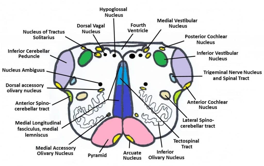
How to draw Medulla Oblongata Crosssection ? Epomedicine
The medulla is divided into two main parts: Place the circle on the bottom line of the oval and draw a line connecting the circle to the oval outline. It connects to the pons superiorly, and to the spinal cord inferiorly. It houses the centers for vital functions of the body, such as those for the heart rate, blood pressure,.
Medulla Oblongata Definition, Structure And Functions
Web medulla oblongata is situated inferior to the pons. The medulla oblongata (medulla) is one of the three regions that make up the brainstem. During development, there’s a disproportion between spinal cord growth and vertebral column growth. Represents medial lemniscus (anteriorly) and medial longitudinal fasciculus (mlf) posteriorly. The pons, the medulla, and the cerebellum.
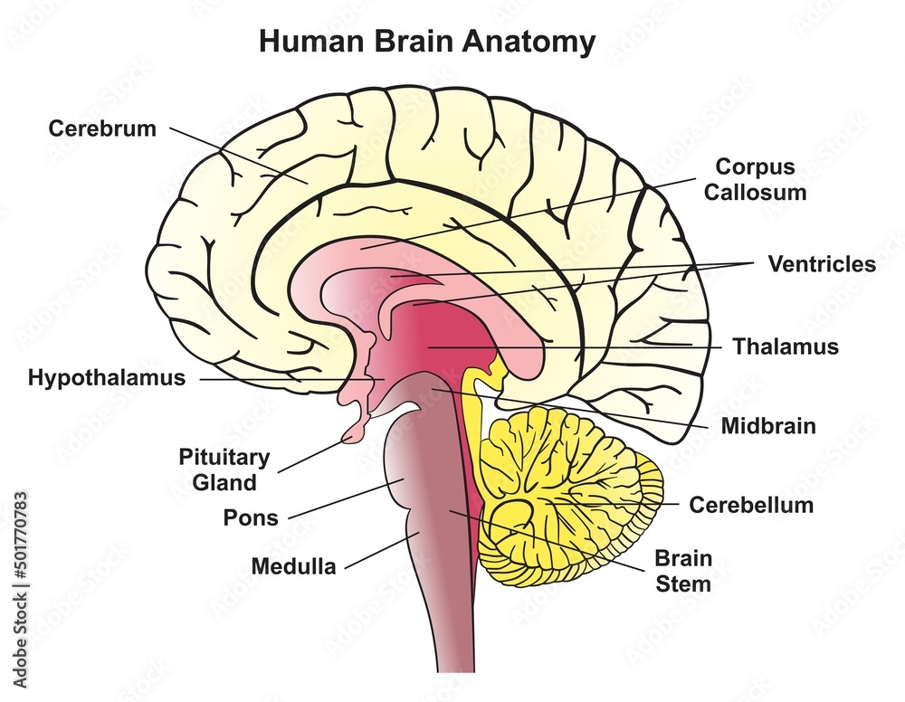
Human brain anatomy infographic diagram showing median section
Web • to begin, draw the cervical spinal cord. • denote that, from a clinician's broad perspective, the medulla is the center for the most basic generators of life. This neuroanatomical atlas is therefore perfectly adapted for the web. Describe the structure of each segment of the nephron. First, draw the different brainstem levels, from superior to inferior:
The Medulla Is Divided Into Two Main Parts:
It houses the centers for vital functions of the body, such as those for the heart rate, blood pressure, and breathing. • denote that, from a clinician's broad perspective, the medulla is the center for the most basic generators of life. Describe the structure of each segment of the nephron. Motor neurons cross from the left.
Their Shape Resembles A Bean, Where We Can Describe The Superior And Inferior Poles, As Well As The Major Convexity Pointed Laterally, And The Minor Concavity Pointed.
The 1 gene with the highest level of enriched expression in pig medulla. Then, erase the rest of the circle. The ventral medulla (the frontal portion) and. So just in case you hear people referring to structures in the brain by these names, that's where those names come from.
Web Just This Part Of The Brain Stem, Up Top.
It is situated inside the vertebral canal of the vertebral column. Medulla and correlate it with functions of both. Sketch a narrow curve above the top line of the brain. Place the circle on the bottom line of the oval and draw a line connecting the circle to the oval outline.
The Renal Medulla Contains The Following Structures:
The pons, the medulla, and the cerebellum. Web it is composed of 64 drawings, illustrations and anatomical charts, all in vector format. And then the hind brain will become the rest of the brain. The medulla oblongata (medulla) is one of the three regions that make up the brainstem.