Metaphase 1 Drawing
Metaphase 1 Drawing - Web in the third step of mitosis, called metaphase, each chromosome lines up in a single file line at the center of the cell. Chromosomes line up at the metaphase plate, under tension from the mitotic spindle. Metaphase i follows prophase i and precedes anaphase i. Web one of the most commonly used diagrams for simplifying meiosis is the metaphase 1 meiosis diagram. Web this is the phase where the two daughter cells produced during the first meiotic division, have their meiotic spindles start to draw the chromosomes to the metaphase plate, again. During prophase i, chromosomes pair up and exchange genetic material, creating more variation. Web the four stages of mitosis are known as prophase, metaphase, anaphase, telophase. This is to prepare the centrosome for division in the next phase. This diagram illustrates the key steps of meiosis at metaphase 1, where homologous chromosomes pair up and align at the center of the cell. Mitosis in a whitefish embryo.
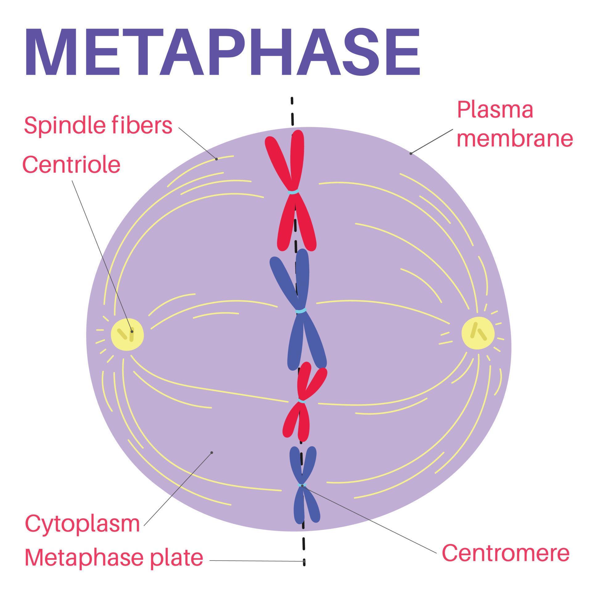
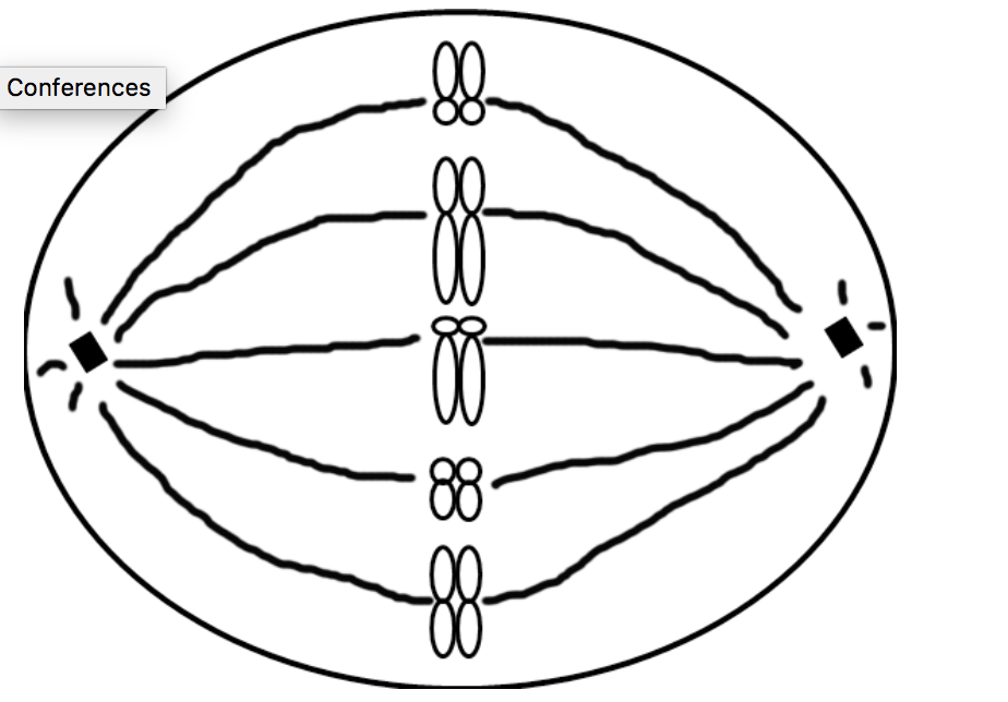
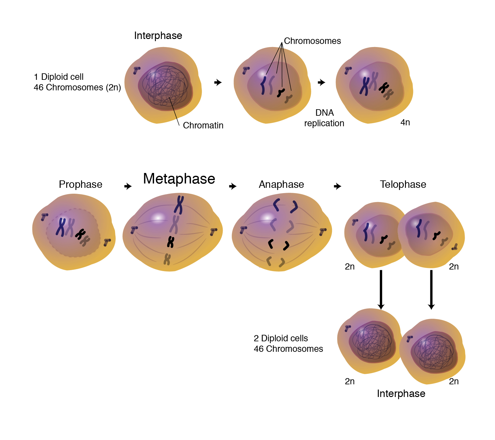
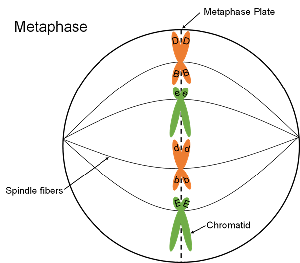
The diagram typically includes labels to denote the chromosomes, spindle fibers, and the centrosomes. Additionally, we’ll mention three other intermediary stages (interphase, prometaphase, and cytokinesis) that play a role in mitosis. Web 3.5k views 3 years ago science diagrams | explained and labelled science diagrams. The g 1 phase (also called the first gap phase) initiates this stage and is focused on cell growth. The fibers arrange the pairs so that homologs are on opposite sides of the metaphase plate (aka equatorial plane). Mitosis in a whitefish embryo. The s phase is next, during which the dna of the chromosomes is replicated. It takes place after a short break known as interkinesis. This diagram illustrates the key steps of meiosis at metaphase 1, where homologous chromosomes pair up and align at the center of the cell. Metaphase of meiosis i occurs when the joined homologous chromosome pairs are moved to the center of the cell by spindle fibers (figure 6).
The phases of meiosis i. Spindle fibers attach to the centromeres of each homologous chromosome. Metaphase i follows prophase i and precedes anaphase i. Web 3.5k views 3 years ago science diagrams | explained and labelled science diagrams. Science > ap®︎/college biology > cell communication and cell cycle > cell cycle. This is the well labelled diagram of. Web in prometaphase i, microtubules attach to the fused kinetochores of homologous chromosomes, and the homologous chromosomes are arranged at the midpoint of the cell in metaphase i. The cell cycle and mitosis: Mitosis, a key part of the cell cycle, involves a series of stages (prophase, metaphase, anaphase, and telophase) that facilitate cell division and genetic information transmission. Web preparation for division happens in three steps:

Metaphase I Biology Dictionary
Web so, during metaphase i, homologue pairs—not individual chromosomes—line up at the metaphase plate for separation. This is the well labelled diagram of. How to draw the metaphase 1 stage of meiosis in exam is the topic. Prophase i, metaphase i, anaphase i, and telophase i. The cell cycle and mitosis:

Cell Division Meiosis HIGH TIME STUDY
Web 3.5k views 3 years ago science diagrams | explained and labelled science diagrams. Science > ap®︎/college biology > cell communication and cell cycle > cell cycle. Mitosis, a key part of the cell cycle, involves a series of stages (prophase, metaphase, anaphase, and telophase) that facilitate cell division and genetic information transmission. It is visibly obvious that replication has.
la métaphase est une étape de la mitose dans le cycle cellulaire
This is to prepare the centrosome for division in the next phase. How to draw the metaphase 1 stage of meiosis in exam is the topic. The cells pinch in the center and divide again.the final outcome is four cells, each with half of the genetic. The phases of meiosis i. The paired chromosomes line up.

FileMitotic Metaphase.svg Wikipedia
During prophase i, chromosomes pair up and exchange genetic material, creating more variation. The cell cycle and mitosis: Metaphase is a stage in eukaryotic cell division in which the chromosomes align on the metaphase plate in the middle of the cell. The diagram typically includes labels to denote the chromosomes, spindle fibers, and the centrosomes. Web preparation for division happens.

Chapter 15 Meiosis and Mitosis Biology 211 with Boury at Iowa State
This diagram illustrates the key steps of meiosis at metaphase 1, where homologous chromosomes pair up and align at the center of the cell. The paired chromosomes line up. Drawing of chromosomes during mitosis by walther flemming, circa 1880. Additionally, we’ll mention three other intermediary stages (interphase, prometaphase, and cytokinesis) that play a role in mitosis. Mitosis in a whitefish.

Metaphase — Definition & Diagrams Expii
Web 3.5k views 3 years ago science diagrams | explained and labelled science diagrams. This is to prepare the centrosome for division in the next phase. Prophase i, metaphase i, anaphase i, and telophase i. In anaphase i, the homologous chromosomes are separated. During prophase i, chromosomes pair up and exchange genetic material, creating more variation.
Solved Draw a metaphase I of meiosis of THIS cell. What
Additionally, we’ll mention three other intermediary stages (interphase, prometaphase, and cytokinesis) that play a role in mitosis. Mitosis in a whitefish embryo. The fibers arrange the pairs so that homologs are on opposite sides of the metaphase plate (aka equatorial plane). During the four phases of mitosis, nuclear division occurs in order for one cell to split into two. The.

Stages Of Metaphase 1
Mitosis in a whitefish embryo. It is the longest phase of meiotic division involving a series of events and is divided into the following steps: The cell cycle and mitosis: Mitosis in a whitefish embryo. The stages of prophase and prometaphase come before metaphase.
Metaphase
Web preparation for division happens in three steps: Metaphase is a stage in eukaryotic cell division in which the chromosomes align on the metaphase plate in the middle of the cell. The fibers arrange the pairs so that homologs are on opposite sides of the metaphase plate (aka equatorial plane). Mitosis in a whitefish embryo. Web this is the phase.
Metaphase Definition and Examples Biology Online Dictionary
How to draw the metaphase 1 stage of meiosis in exam is the topic. The phases of meiosis i. It is the longest phase of meiotic division involving a series of events and is divided into the following steps: Web in prometaphase i, microtubules attach to the fused kinetochores of homologous chromosomes, and the homologous chromosomes are arranged at the.
Metaphase I Follows Prophase I And Precedes Anaphase I.
In meiosis i, cells go through four phases: Spindle fibers attach to the centromeres of each homologous chromosome. During prophase i, chromosomes pair up and exchange genetic material, creating more variation. The s phase is next, during which the dna of the chromosomes is replicated.
The Two Sister Chromatids Of Each Chromosome Are Captured By Microtubules From Opposite Spindle Poles.
Phase, also called the first gap phase, the cell grows physically larger, copies organelles, and makes the molecular building blocks it will need in later steps. In metaphase i, chromosomes line up in the middle of the cell. Web the four stages of mitosis are known as prophase, metaphase, anaphase, telophase. The phases of meiosis i.
The G 1 Phase (Also Called The First Gap Phase) Initiates This Stage And Is Focused On Cell Growth.
Web so, during metaphase i, homologue pairs—not individual chromosomes—line up at the metaphase plate for separation. Web the first metaphase of meisosis i encompasses the alignment of paired chromosomes along the center (metaphase plate) of a cell, ensuring that two complete copies of chromosomes are present in the resulting two daughter cells of meiosis i. Metaphase is a stage in eukaryotic cell division in which the chromosomes align on the metaphase plate in the middle of the cell. Chromosomes line up at the metaphase plate, under tension from the mitotic spindle.
Furthermore, Cells Can Be Experimentally Arrested At Metaphase With Mitotic.
Web in prometaphase i, microtubules attach to the fused kinetochores of homologous chromosomes, and the homologous chromosomes are arranged at the midpoint of the cell in metaphase i. The starting cell is diploid, 2n = 4. Prophase i, metaphase i, anaphase i, and telophase i. It is visibly obvious that replication has not occurred.