Microscope Drawing And Parts
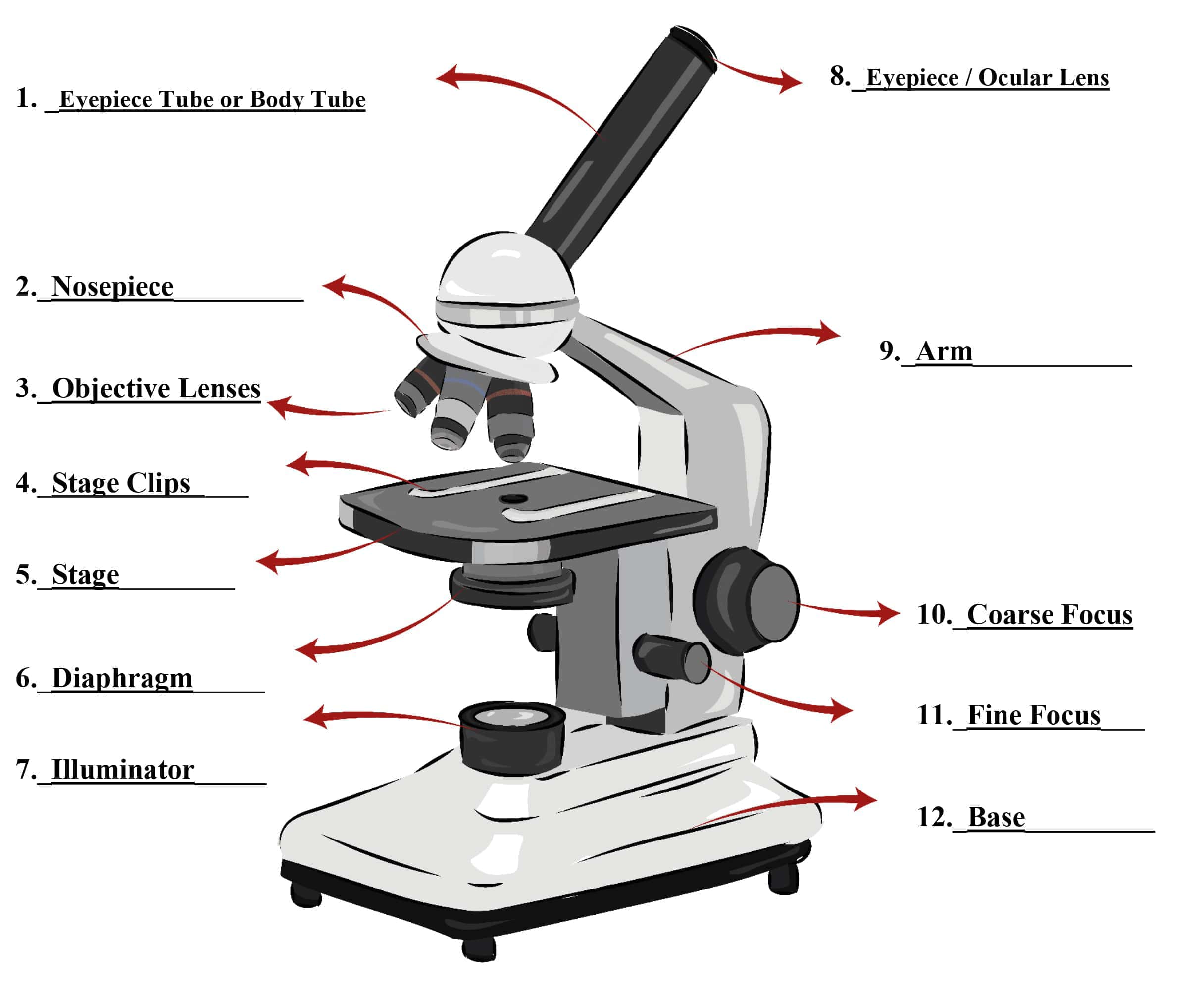
Microscope Drawing And Parts - Use a curved line to enclose a rounded shape beneath the head. The ocular lens is the lens close to the eye, and the objective lens is the lens close to the object. First, the purpose of a microscope is to. It is also called a body tube or eyepiece tube. This forms the arm of the microscope. Below this, draw another curved line, leaving the shape open on one side. Connect them at the bottom using curved lines. So, something that was 1 mm wide in real life would be 400 mm wide in the microscope image. Power = 10 x 4 = 40 power = 10 x 10 = 100 power = 10 x 40 = 400 what happens as the power of magnification increases? It is to be noted that.
This forms the arm of the microscope. Web parts of a microscope. Diagram of parts of a microscope. Learn about the types, parts, history, diagram, and facts of microscope from britannica, the trusted source of knowledge. The base is attached to a frame (arm) that is connected to the head of the device.the base of the microscope provides stability to the device and allows the user’s. In this interactive, you can label the different parts of a microscope. Knobs (fine and coarse) by adjusting the knob, you can adjust the focus of the microscope. Web parts of the optical parts are as follows: Scanning objective lens (4x) low power objective (10x) high power objective lens (40x) oil immersion objective lens (100x) specialty objective lenses. Web the magnification power of a simple microscope is expressed as:
Web parts of a microscope. Web magnification is a measure of how much larger a microscope (or set of lenses within a microscope) causes an object to appear. Web optical components of a compound microscope. Web microscope parts and functions with labeled diagram and functions how does a compound microscope work?. Eyepieces typically have a magnification between 5x & 30x. The eyepiece (or ocular lens) is the lens part at the top of a microscope that the viewer looks through. The magnification power of a simple microscope is about 10, meaning that the specimen. Web the magnification power of a simple microscope is expressed as: It is also called a body tube or eyepiece tube. Web parts of a microscope.
Simple Microscope Definition, Principle, Parts, And Uses » Microscope Club
Use a curved line to enclose a rounded shape beneath the head. The upper part of the microscope that houses the optical elements of the unit.; Supports the microscope head and attaches it to the base. The standard eyepiece has a magnification of 10x. Web optical parts of a compound microscope.
Parts of a Compound Microscope Labeled (with diagrams) Medical
A steady light source (110 volts) used in place of a mirror. The standard eyepiece has a magnification of 10x. It is also called a body tube or eyepiece tube. Diagram of parts of a microscope. If you want to redo an answer, click on the.

Simple Microscope Definition, Principle, Magnification, Parts
Web optical components of a compound microscope. For instance, the light microscopes typically used in high schools and colleges magnify up to about 400 times actual size. Web magnification is a measure of how much larger a microscope (or set of lenses within a microscope) causes an object to appear. The magnification power of a simple microscope is about 10,.

Microscope Diagram Labeled, Unlabeled and Blank Parts of a Microscope
Web parts of the optical parts are as follows: The eyepiece (or ocular lens) is the lens part at the top of a microscope that the viewer looks through. Web the microscope illustrated in figure 5 below was manufactured by hugh powell and peter lealand around 1850. It is also called a body tube or eyepiece tube. Structural support that.
Parts of a Microscope Microscope Parts and Functions Labkafe
It is because it contains two types of lenses; The eyepiece, also known as the “ocular”, is the first magnification lens you will look through in a compound microscope. Web the magnification power of a simple microscope is expressed as: Web the microscope illustrated in figure 5 below was manufactured by hugh powell and peter lealand around 1850. The evolution.

Microscope Diagram Labeled, Unlabeled and Blank Parts of a Microscope
The ocular lens is the lens close to the eye, and the objective lens is the lens close to the object. Supports the tube and connects it to the base. Web the common light microscope used in the laboratory is called a compound microscope. Web microscope is an instrument that magnifies small objects and reveals their details. Use this with.
Parts Of A Microscope With Functions And Labeled Diagram Images
The magnification power of a simple microscope is about 10, meaning that the specimen. Learn about the types, parts, history, diagram, and facts of microscope from britannica, the trusted source of knowledge. If your microscope has a mirror, it is used to reflect light from an external light source up through the bottom. But small businesses lag behind large companies.
Parts of a Microscope SmartSchool Systems
Use a curved line to enclose a rounded shape beneath the head. Fully open field and condenser diaphragms and focus on specimen using x10 objective. Web magnification is a measure of how much larger a microscope (or set of lenses within a microscope) causes an object to appear. The eyepiece (or ocular lens) is the lens part at the top.

Parts of a microscope with functions and labeled diagram (2023)
The bottom of the microscope, used for support. Parts of a powell and leland microscope diagram Connects the eyepiece to the objective lenses. Use screws at front of condenser to centre field diaphragm and open field diaphragm to fill view. Drag and drop the text labels onto the microscope diagram.

Simple Microscope Drawing With Parts Micropedia
Web eyepiece (ocular lens) with or without pointer: Web the web page titled “parts of a microscope with labeled diagram and functions” has the following key takeaways: The ocular lens is the lens close to the eye, and the objective lens is the lens close to the object. Many optical parts of a microscope work together to magnify and produce.
Answers Pdf Printable Version Here.
Many optical parts of a microscope work together to magnify and produce an image of the specimen placed on a slide. Parts of a powell and leland microscope diagram Use this with the microscope parts activity to help students identify and label the main parts of a microscope and then describe their functions. The majority of the microscope models today have the knobs mounted on the same part of the device.
It Is Because It Contains Two Types Of Lenses;
The evolution of the microbiology field put to perspective the need to identify, view, observe and understand microorganisms, including their structural morphologies and mechanisms. Web magnification is a measure of how much larger a microscope (or set of lenses within a microscope) causes an object to appear. Web the common light microscope used in the laboratory is called a compound microscope. Download the label the parts of the microscope:
The Eyepiece, Also Known As The “Ocular”, Is The First Magnification Lens You Will Look Through In A Compound Microscope.
Scanning objective lens (4x) low power objective (10x) high power objective lens (40x) oil immersion objective lens (100x) specialty objective lenses. Microbiology’s scope is to study organisms and minute. Connects the eyepiece to the objective lenses. Web optical parts of a compound microscope.
F = Focal Length Of The Convex Lens.
Learn about the types, parts, history, diagram, and facts of microscope from britannica, the trusted source of knowledge. But small businesses lag behind large companies on productivity. Use screws at front of condenser to centre field diaphragm and open field diaphragm to fill view. 800.942.0528 (us toll free) 1.760.438.0528 (international) microscope world explains the parts of the microscope, including a printable worksheet for schools and home.