Pineal Gland Drawing
Pineal Gland Drawing - Replaced by connective tissue after puberty. Depictions of pine cones and sacred eyes have been linked to this mysterious biological feature and can be found in ancient cultures around the world. The pineal gland, conarium, or epiphysis cerebri. Web glands and organs of the endocrine system; Also visible is a corpus arenaceum. Higher magnification of the pineal gland shows pinealocytes arranged in poorly defined lobules. 1 since the advent of mr brain imaging, the incidence of pineal cysts has been reported to vary from 0.58% to 10.8% in large consecutive brain mr imaging studies. The shape of the gland resembles a pine cone, which gives it its name. Also called epiphysis, pineal body. Your pineal gland’s main job is to help control the circadian cycle of sleep and wakefulness by secreting melatonin.
These cells produce and secrete the hormone melatonin in response to low light levels. Pineal gland pituitary gland parathyrad gland 4. Web the pineal gland is small glandular body, approximately 6mm long. Depictions of pine cones and sacred eyes have been linked to this mysterious biological feature and can be found in ancient cultures around the world. Your pineal gland’s main job is to help control the circadian cycle of sleep and wakefulness by secreting melatonin. It is shaped like a pine cone, from which its name is derived. Through diverse techniques, they highlighted its. Connective tissue septa, extending from the pia mater capsule, penetrate the parenchyma of the gland. Between superior colliculi at base of brain; Up to 23% in healthy.
The shape of the gland resembles a pine cone, which gives it its name. The pineal gland or pineal body is a small gland in the middle of the head. There are more than 99,000 vectors, stock photos & psd files. Cut outs | vectors | black & white. Web accessed may 11th, 2024. The pineal gland, conarium, or epiphysis cerebri. Between superior colliculi at base of brain; Produces melatonin, which helps regulate circadian rhythms. Diagram of pituitary and pineal glands in the human brain. It is made up of pinealocytes.

Pineal gland, illustration Stock Image F036/1604 Science Photo
Higher magnification of the pineal gland shows pinealocytes arranged in poorly defined lobules. Huge collection, amazing choice, 100+ million high quality, affordable rf and rm images. Connective tissue septa, extending from the pia mater capsule, penetrate the parenchyma of the gland. Pineal gland pituitary gland parathyrad gland 4. Up to 23% in healthy.

Pineal gland, illustration Stock Image F036/1617 Science Photo
See pineal gland stock video clips. High blood levels of melatonin induce drowsiness. Web download scientific diagram | e histological picture of the pineal gland in high magnification (3100) showing pinealocytes arranged in cords and blood capillaries. Between superior colliculi at base of brain; There is an enigmatic gland which seems to be hidden away in the human brain that.

Pineal gland anatomical cross section vector illustration diagram with
Up to 23% in healthy. There are more than 99,000 vectors, stock photos & psd files. Diagram of pituitary and pineal glands in the human brain. Also called epiphysis, pineal body. Web pineal gland drawing stock photos and images.

Pineal gland, illustration Stock Image F036/1621 Science Photo
Connective tissue septa penetrate the gland, subdividing it into indistinct lobules. Web the pineal gland is small glandular body, approximately 6mm long. Web the pineal gland (also known as the pineal body or epiphysis cerebri) is a small endocrine gland in the brain of most vertebrates. See pineal gland stock video clips. Huge collection, amazing choice, 100+ million high quality,.

Pineal gland, illustration Stock Image F036/1616 Science Photo
Web accessed may 11th, 2024. Web glands and organs of the endocrine system; Drawing showing the anatomy of the pineal gland and pituitary gland in the brain. No need to register, buy now! Connective tissue septa, extending from the pia mater capsule, penetrate the parenchyma of the gland.

Pineal gland, illustration Stock Photo Alamy
The pineal gland has a predilection for calcification which is invariably histologically present in adults but rarely seen below the age of 10 years 6. The pineal gland or pineal body is a small gland in the middle of the head. Pineal gland pituitary gland parathyrad gland 4. No need to register, buy now! Up to 23% in healthy.
Pineal Gland & its Function Cyst & Calcified Pineal Gland
The shape of the gland resembles a pine cone, which gives it its name. There are more than 99,000 vectors, stock photos & psd files. Through diverse techniques, they highlighted its. What is the pineal gland? Web accessed may 11th, 2024.

Pineal gland, illustration Stock Image F036/1620 Science Photo
Connective tissue septa penetrate the gland, subdividing it into indistinct lobules. Also called epiphysis, pineal body. The gland is surrounded by a capsule of pia mater and arachnoid elements; The pineal gland or pineal body is a small gland in the middle of the head. Between superior colliculi at base of brain;

Pineal gland, illustration Stock Image F036/1618 Science Photo
No need to register, buy now! Web the pineal gland is an endocrine structure of the diencephalon of the brain, and is located inferior and posterior to the thalamus. Replaced by connective tissue after puberty. There are two types of cells present within the gland: Web download scientific diagram | e histological picture of the pineal gland in high magnification.
Pineal gland Anatomy, histology and blood supply Kenhub
Higher magnification of the pineal gland shows pinealocytes arranged in poorly defined lobules. Web download scientific diagram | e histological picture of the pineal gland in high magnification (3100) showing pinealocytes arranged in cords and blood capillaries. 3.2k views 3 years ago endocrine gland histology. These cells produce and secrete the hormone melatonin in response to low light levels. Web.
All Images Photos Vectors Illustrations 3D Objects.
The pineal gland consists of portions of neurons, neuroglial cells, and. 3.2k views 3 years ago endocrine gland histology. Histology of pineal gland explanation with step by step drawing, lecture on pineal gland | anatomy | practical | journal drawing |. Web accessed may 11th, 2024.
The Pineal Gland, Conarium, Or Epiphysis Cerebri.
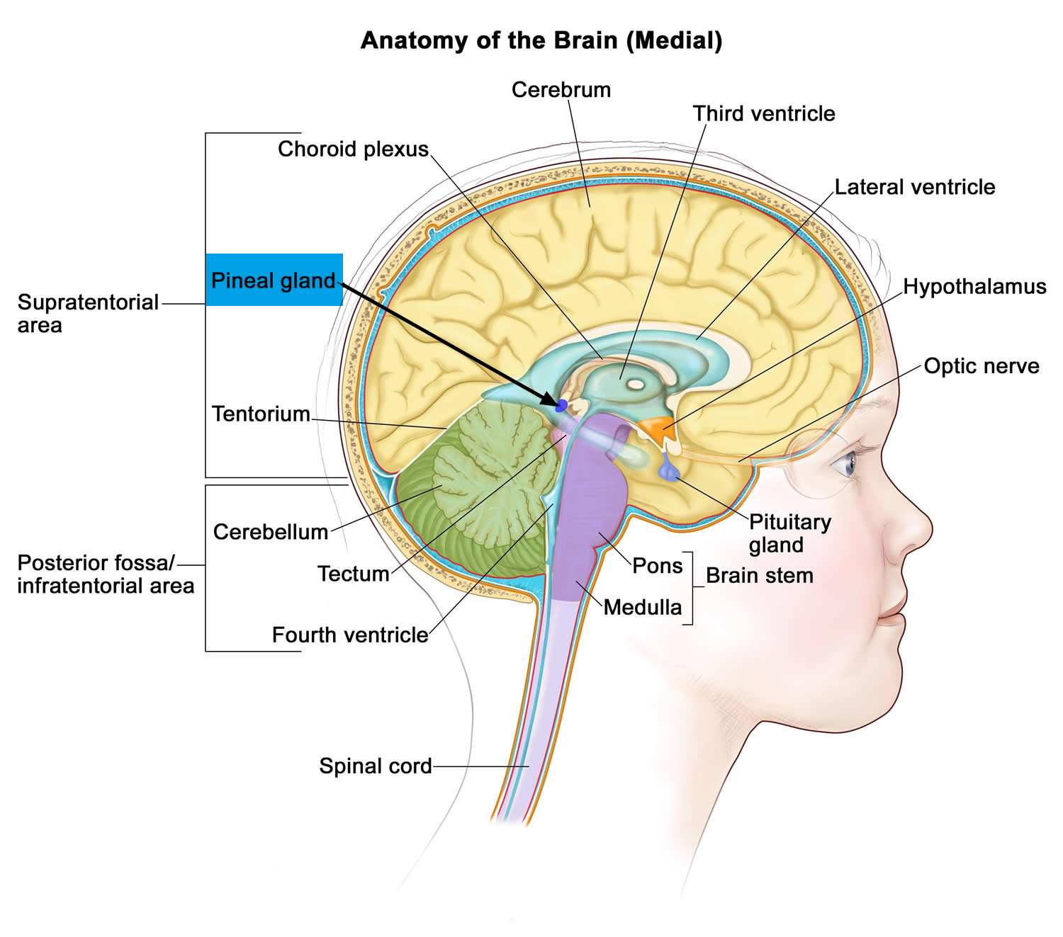
Drawing shows the hypothalamus, pituitary gland, pineal gland, thyroid gland, thymus, adrenal gland, pancreas, ovaries (female), and testes (male). It is made up of pinealocytes. Web artists used paintings and drawings as a canvas to explore both the symbolic significance and anatomical representation of the pineal gland. Web the anatomy of the pineal gland, along with the pituitary gland, is displayed in the image below.
Higher Magnification Of The Pineal Gland Shows Pinealocytes Arranged In Poorly Defined Lobules.
Between superior colliculi at base of brain; Through diverse techniques, they highlighted its. The gland is surrounded by a capsule of pia mater and arachnoid elements; Web the pineal gland (also known as the pineal body or epiphysis cerebri) is a small endocrine gland in the brain of most vertebrates.
No Need To Register, Buy Now!
Web pineal gland drawing stock photos and images. Web find the perfect pineal gland drawing image. It is a neuroendocrine gland that secretes the hormone melatonin and several other polypeptide hormones that have. Develops at month 2 of gestation as diverticulum in diencephalic roof of third ventricle.