Prophase Drawing
Prophase Drawing - All other use is by permission only. Prophase i, metaphase i, anaphase i, and telophase i. Cytokinesis, the process of cell division, occurs during the last stage of mitosis (telophase). You can learn more about these stages in the video on mitosis. Prophase, metaphase, anaphase, and telophase. Prophase, metaphase, anaphase, and telophase. Edupic graphical resource is a teacher designed free image resource for use by teachers and students. In cytokinesis, the cytoplasm of. Humans have 46) but the diagrams below show mitosis of an animal cell with only four chromosomes, for simplicity. Prophase is the starting stage of cell division in eukaryotes.
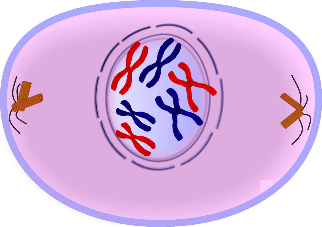
Microtubules align chromosomes along metaphase plate. Prophase i, metaphase i, anaphase i, and telophase i. If a nucleolus is still present or you can distinguish a forming spindle, label these as well. Sometimes a cell in g 0 will move back into g 1 and continue. Prophase is the first phase of mitosis. Prophase, prometaphase, metaphase, anaphase, and telophase. Web in meiosis i, cells go through four phases: Humans have 46) but the diagrams below show mitosis of an animal cell with only four chromosomes, for simplicity. By the end of this section, you will be able to: Web prophase → metaphase → anaphase → telophase.
Kinetochore microtubules shorten, pulling sister chromatids to opposite poles, polar microtubules elongate, lengthening dividing cell. Label the cell wall, plasma membrane, nuclear envelope (or where it would be), and chromosomes. This part of mitosis is all about preparing. In each round of division, cells go through four stages: All images contained within are free for use by educational professionals and the students they serve without permission. Some textbooks list five, breaking prophase into an early phase (called prophase) and a late phase (called prometaphase). These phases are prophase, prometaphase, metaphase, anaphase, and telophase. Prophase i, metaphase i, anaphase i, and telophase i. Mitosis, a key part of the cell cycle, involves a series of stages (prophase, metaphase, anaphase, and telophase) that facilitate cell division and genetic information transmission. Web today, mitosis is understood to involve five phases, based on the physical state of the chromosomes and spindle.

Biology 2e, The Cell, Cell Reproduction, The Cell Cycle OpenEd CUNY
In cytokinesis, the cytoplasm of. Kinetochore microtubules shorten, pulling sister chromatids to opposite poles, polar microtubules elongate, lengthening dividing cell. Humans have 46) but the diagrams below show mitosis of an animal cell with only four chromosomes, for simplicity. Web since cell division occurs twice during meiosis, one starting cell can produce four gametes (eggs or sperm). Prophase i is.

Prophase Tutorial Sophia Learning
These phases are prophase, prometaphase, metaphase, anaphase, and telophase. Web since cell division occurs twice during meiosis, one starting cell can produce four gametes (eggs or sperm). Prophase, metaphase, anaphase, and telophase. Discuss the behavior of chromosomes during mitosis and how the cytoplasmic content divides during cytokinesis. Prophase, in both mitosis and meiosis, is recognized by the condensing of chromosomes.
Prophase Diagrams
Web here is a diagram of what a nematode cell nucleus looks like after prophase and metaphase. During interphase, the parent cell’s chromosomes are replicated, but they aren’t yet visible. Some textbooks list five, breaking prophase into an early phase (called prophase) and a late phase (called prometaphase). Microtubules align chromosomes along metaphase plate. In each round of division, cells.
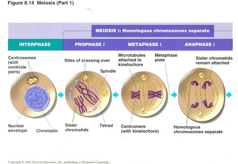
Diagram Of Prophase
Prophase, metaphase, anaphase, and telophase. By the end of this section, you will be able to: Prophase, metaphase, anaphase, and telophase. Prophase i, metaphase i, anaphase i, and telophase i. Microtubules align chromosomes along metaphase plate.
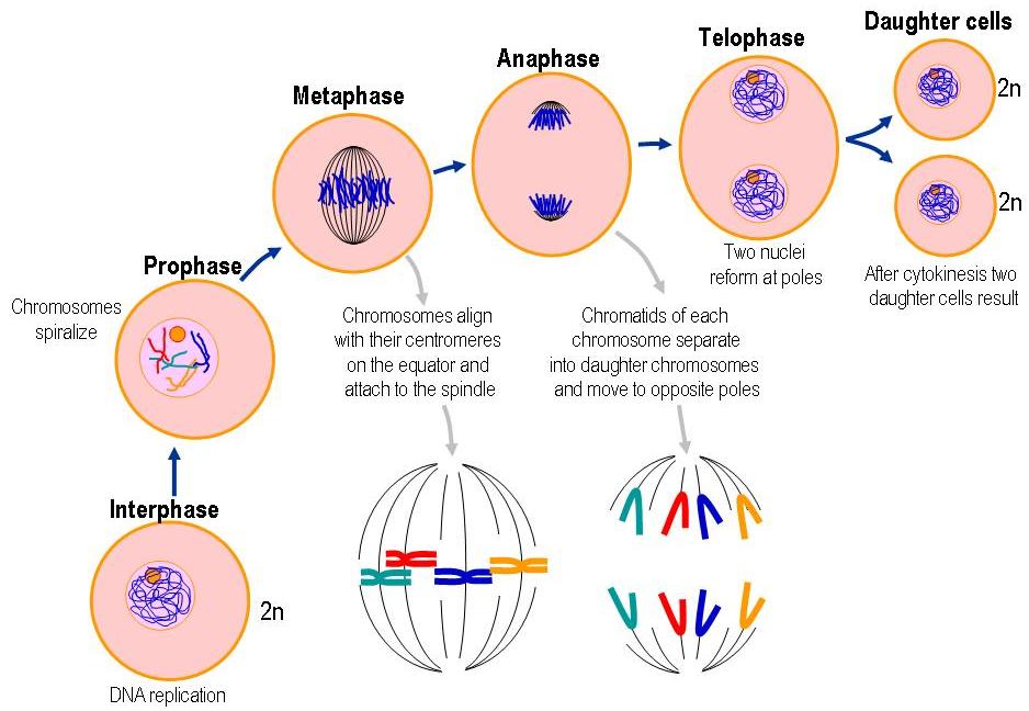
Diagram Of Prophase
Web mitosis consists of four basic phases: You may find that some accounts of mitosis further subdivide the process to include prometaphase between prophase and metaphase. Web in meiosis i, cells go through four phases: By the end of this section, you will be able to: During interphase, the parent cell’s chromosomes are replicated, but they aren’t yet visible.

Prophase. The First Stage of Mitosis 12682013 Vector Art at Vecteezy
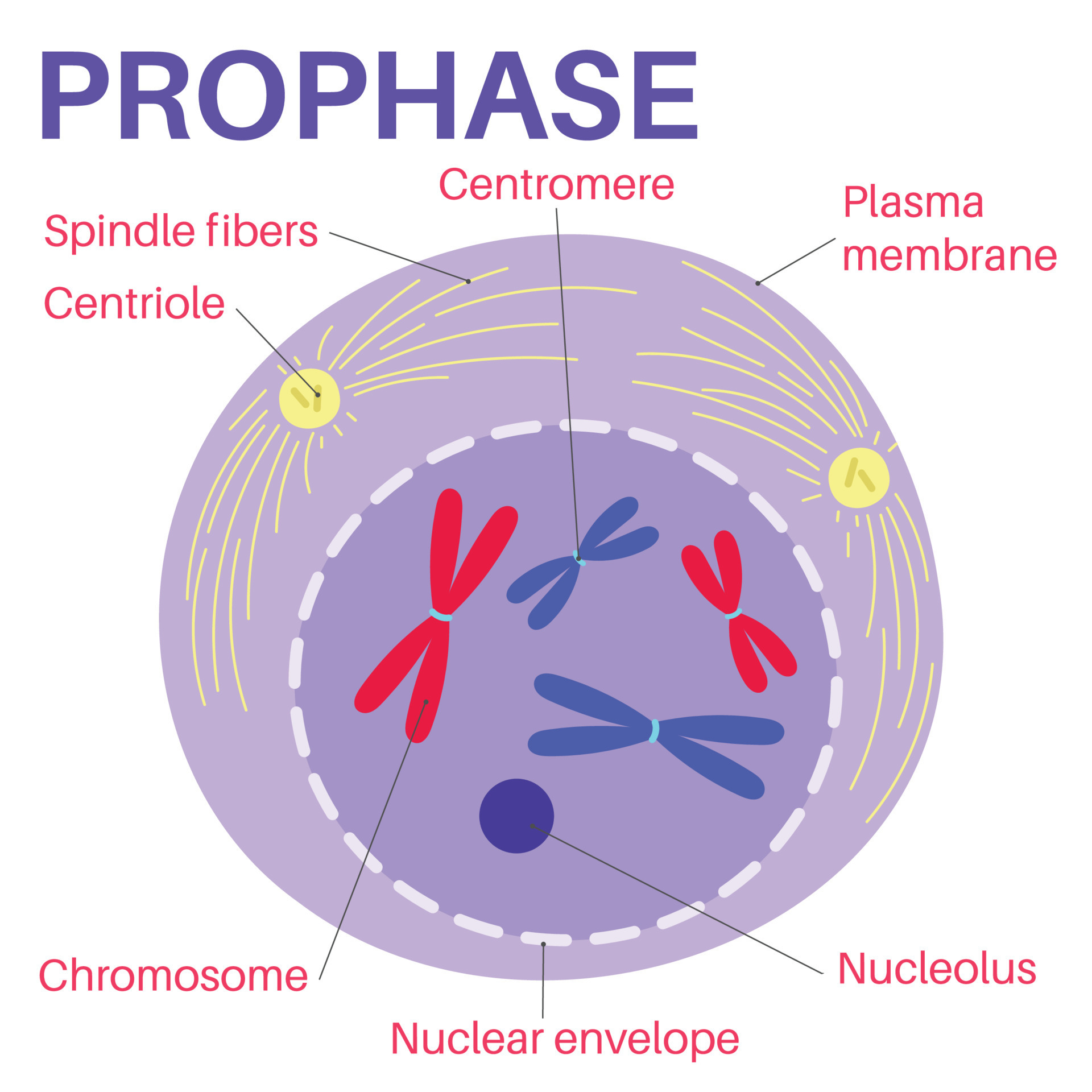
Centrosomes and microtubules play pivotal roles in orchestrating this complex process, ensuring the successful replication of cells. If a nucleolus is still present or you can distinguish a forming spindle, label these as well. Prophase i, metaphase i, anaphase i, and telophase i. Kinetochore microtubules shorten, pulling sister chromatids to opposite poles, polar microtubules elongate, lengthening dividing cell. Web prophase.
Prophase is the first stage of cell division. 14268877 Vector Art at
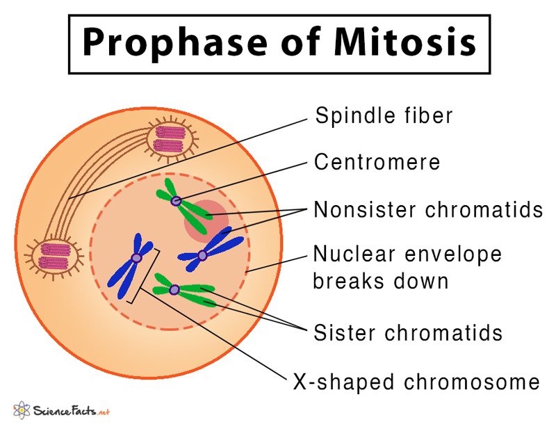
This is when the genetic fibers within the cell’s nucleus, known as chromatin, begin to condense and become tightly compacted together. In this case, these cells move from g 1 of the cell cycle into a resting phase known as g 0. In this exercise, we will consider prometaphase a component of prophase. This part of mitosis is all about.
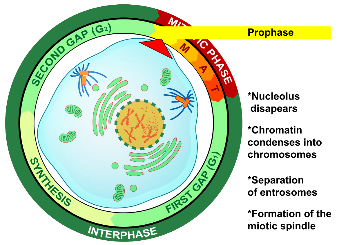
Diagram Of Prophase
Web draw an onion cell in prophase. Prophase is the starting stage of cell division in eukaryotes. Humans have 46) but the diagrams below show mitosis of an animal cell with only four chromosomes, for simplicity. Most organisms contain many chromosomes in the nuclei of their cells (eg. Label the cell wall, plasma membrane, nuclear envelope (or where it would.
Mitosis Definition, Stages, & Purpose, with Diagram
If a nucleolus is still present or you can distinguish a forming spindle, label these as well. Define the quiescent g 0 phase. By the end of this section, you will be able to: Humans have 46) but the diagrams below show mitosis of an animal cell with only four chromosomes, for simplicity. Edupic graphical resource is a teacher designed.
EduPic Cell Drawings
Web here is a diagram of what a nematode cell nucleus looks like after prophase and metaphase. In the fourth step, anaphase , the mitotic spindles pry each chromatid apart from its copy, and drag them to the opposite side of the cell. If a nucleolus is still present or you can distinguish a forming spindle, label these as well..
Mitosis, A Key Part Of The Cell Cycle, Involves A Series Of Stages (Prophase, Metaphase, Anaphase, And Telophase) That Facilitate Cell Division And Genetic Information Transmission.
Some textbooks list five, breaking prophase into an early phase (called prophase) and a late phase (called prometaphase). Prophase is the first phase of mitosis. In cytokinesis, the cytoplasm of. These phases are prophase, prometaphase, metaphase, anaphase, and telophase.
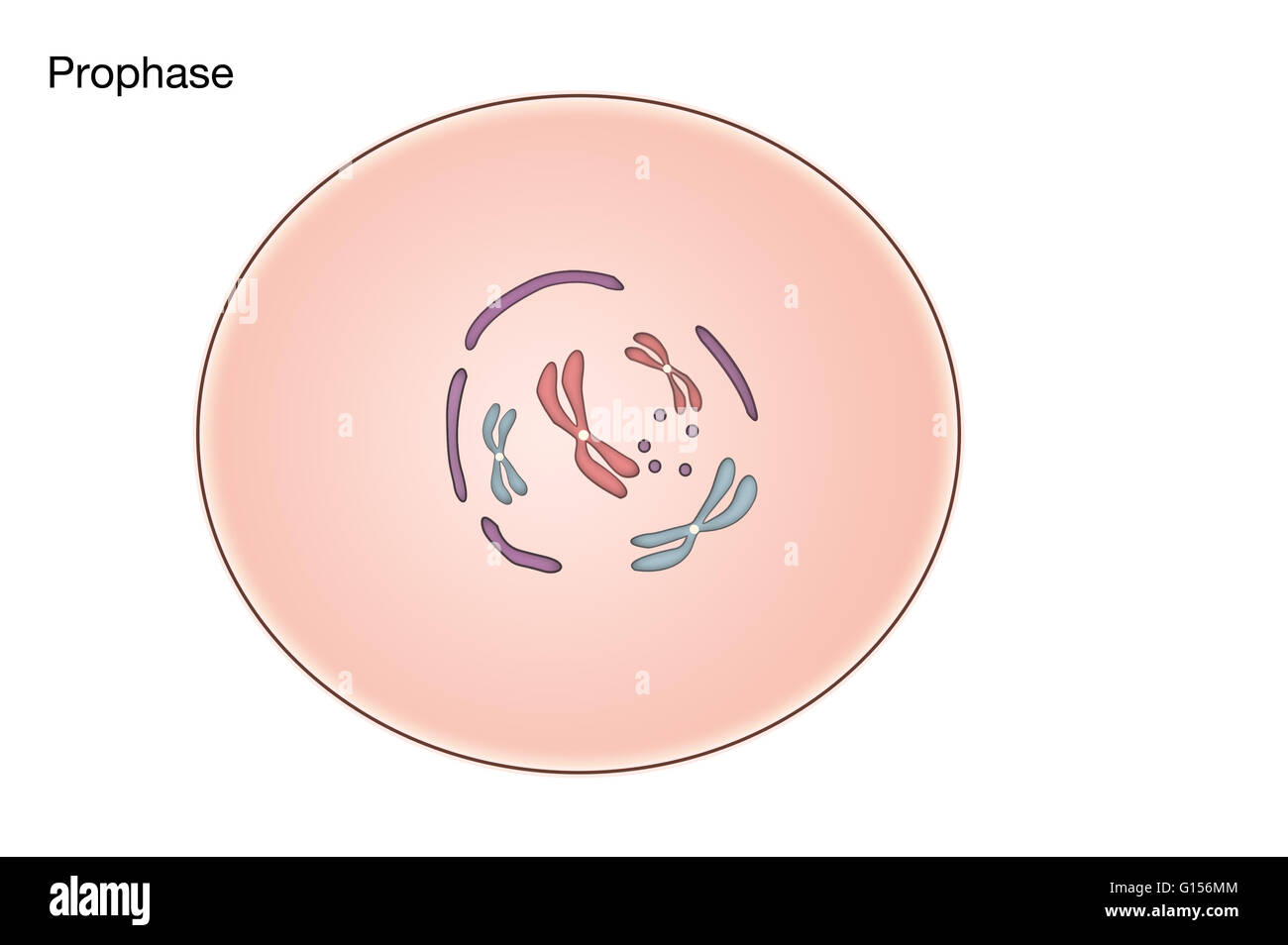
Prophase, In Both Mitosis And Meiosis, Is Recognized By The Condensing Of Chromosomes And Separation Of The Centrioles In The Centrosome.
Sometimes a cell in g 0 will move back into g 1 and continue. All other use is by permission only. Beginning after interphase, dna has already been replicated when the cell enters prophase. Web mitosis consists of five morphologically distinct phases:
In This Phase, The Chromosomes Consist Of Two Identical Chromatids Called Sister Chromatids.
During this phase, the dna forms into chromosomes, which we can actually see. Define the quiescent g 0 phase. Edupic graphical resource is a teacher designed free image resource for use by teachers and students. Prophase, metaphase, anaphase, and telophase.
You Can Learn More About These Stages In The Video On Mitosis.
Web prophase → metaphase → anaphase → telophase. Nuclear membrane breaks down, chromatin condenses, mitotic spindle forms and attaches to kinetochores. Web draw an onion cell in prophase. Web here is a diagram of what a nematode cell nucleus looks like after prophase and metaphase.