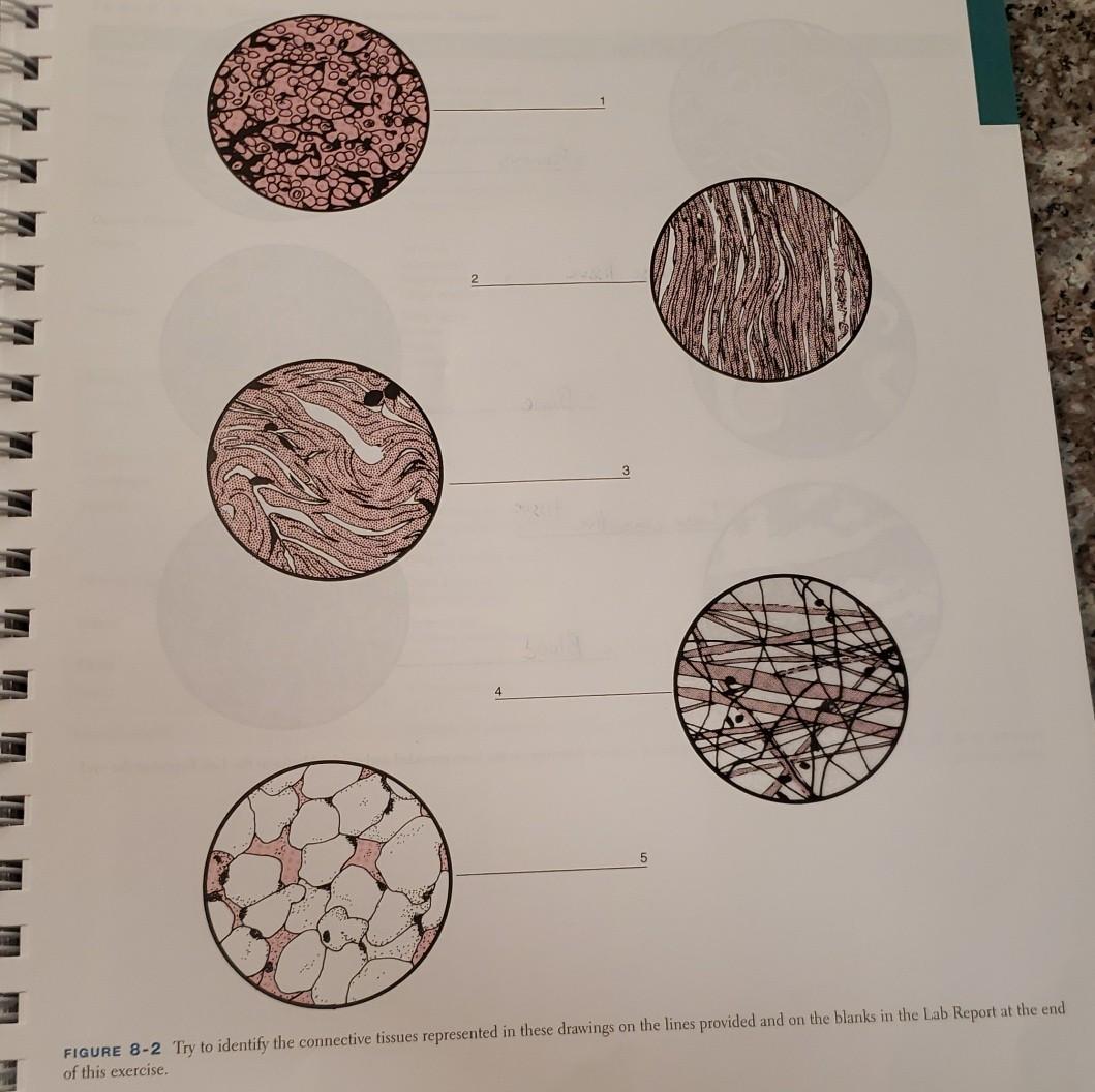
Reticular Connective Tissue Drawing
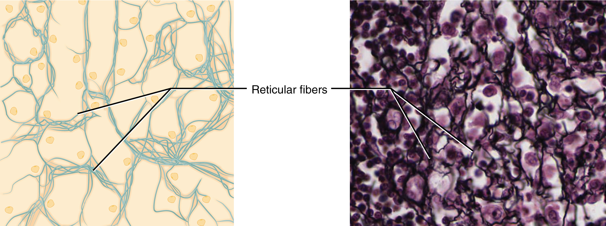
Reticular Connective Tissue Drawing - The cells that make the reticular fibers are fibroblasts called reticular cells. The fibers form attachment sites for lymphocytes and other immune cells. Obtain a slide of a spleen or lymph node with reticular connective tissue from the slide box. Recognize interstitial (fibrillar) collagens and elastic fibers at the light and em level. Supports other cell types, including white blood cells mast cells, and macrophages. Web reticular tissue, a form of loose connective tissue wherein reticular fibres are the most predominant fibrous constituent, serves as the supporting structure of the bone marrow, liver and lymphoid organs (spleen, lymph nodes, and tonsils). However connective tissue differs from other types in that its cells are loosely, rather than tightly, packed. Draw and label reticular tissue: Forms stroma of liver, spleen, bone marrow, and lymph. Web reticular connective tissue 10x.
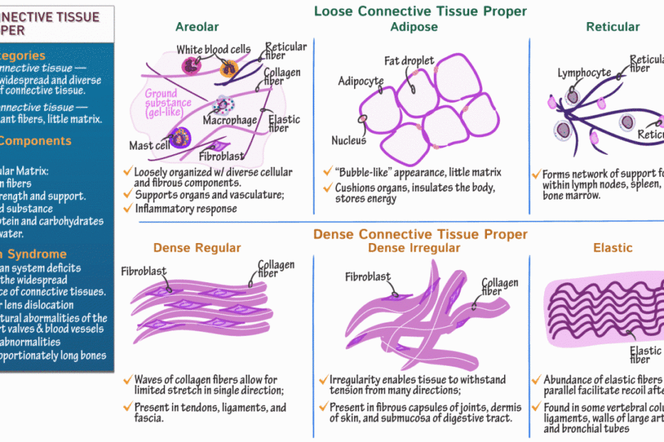
They are present in relatively high. Reticular connective tissue forms a scaffolding for other cells in several organs, such as lymph nodes and bone marrow. Web connective tissue is the tissue that connects or separates, and supports all the other types of tissues in the body. Web the reticular connective tissue network provides structure within solid organs and soft tissues so it is found in locations such as the spleen, the lymphatic system, the liver, and bone marrow which in adults is found primarily in the axial skeleton. Web reticular connective tissue. Summary of the properties of the major types of connective tissue proper. Web reticular connective tissue is a type of connective tissue with a network of reticular fibers, made of type iii collagen (reticulum = net or network). Forms stroma of liver, spleen, bone marrow, and lymph. This special connective tissue forms the stroma for hemopoietic tissues and lymphoid structures and organs, except the thymus. A slide of reticular connective tissue from a human spleen.
Reticular tissue is a special subtype of connective tissue that is indistinguishable during routine histological staining. Learn everything about it in the f. Web reticular connective tissue, 40x. Reticular fibers form the stroma. Web the outer portion of the lymph node contains aggregations of lymphocytes organized in. Fine reticular fibers stain faintly; Is a fine interlacing network of reticular fibers and reticular cells. *font labels changed to red for easier visualization because the slide was stained dark. Web reticular connective tissue. A slide of reticular connective tissue from a human spleen.
Reticular Connective Tissue Drawing Master the Art of Illustrating
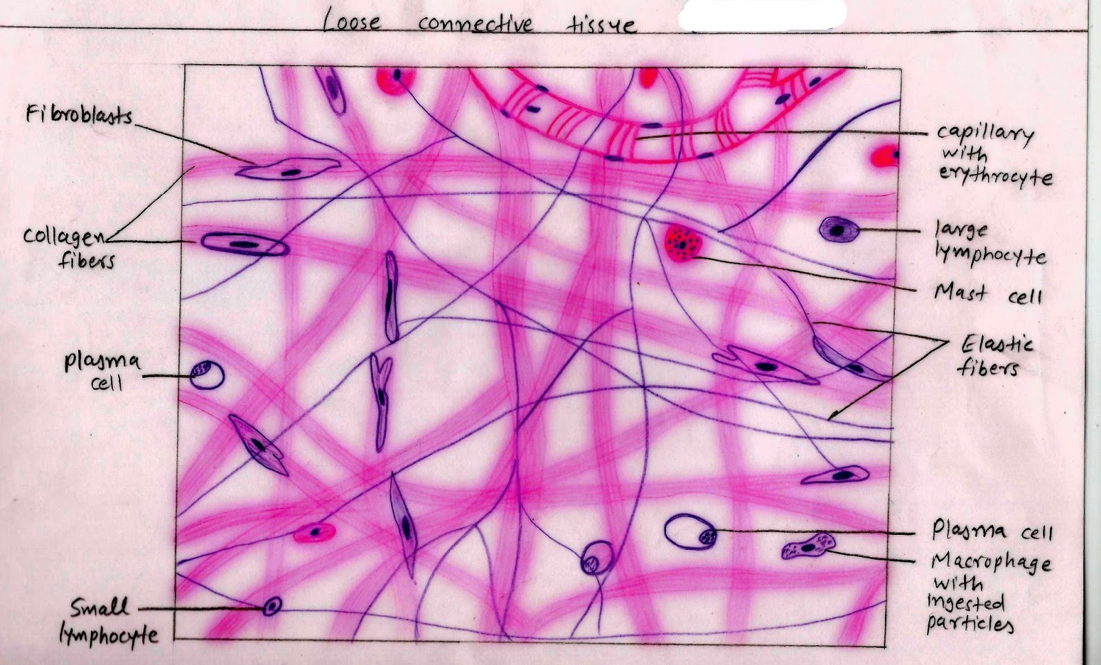
A slide of reticular connective tissue from a human spleen. In drawing images of connective tissue proper preparations seen under the microscope, it is important to simplify the visuals. Web the outer portion of the lymph node contains aggregations of lymphocytes organized in. Reticular tissue forms the stroma for the spleen, lymph. Reticular connective tissue is named for the reticular.

Reticular Connective Tissue, 40X Histology
Forms stroma of liver, spleen, bone marrow, and lymph. Reticular fibers are synthesized by special fibroblasts called reticular cells.the fibers are thin. Web reticular connective tissue 40x. Web the tissue structure looks quite similar to that of elastic connective tissue. Recognize interstitial (fibrillar) collagens and elastic fibers at the light and em level.

Reticular Connective Tissue Labeled
Web reticular connective tissue. Lymphoid organs (lymph nodes, bone marrow, and spleen) function of reticular connective tissue. Web describe the functions of cells commonly found in connective tissue and be able to identify them at the light and em microscope level. This is the most easily recognized tissue and will be found widely distributed in every organ. Web the tissue.
Reticular Connective Tissue Drawing Master the Art of Illustrating
Reticular connective tissue is named for the reticular fibers which are the main structural part of the tissue. Web reticular connective tissue is composed of a meshwork of reticular fibers (type iii collagen) in an open arrangement. Reticular fibers (type iii collagen) stained black surround each nodule. Supports other cell types, including white blood cells mast cells, and macrophages. Web.

Mammalian Tissues Lab Notebook Students Coursework
The structural framework of collagen lattice in reticular tissues provides great strength and support to the organs of the human body system. May anchor to collagenous septa, which divide organs into lobes. Web the reticular connective tissue network provides structure within solid organs and soft tissues so it is found in locations such as the spleen, the lymphatic system, the.

chapter 4 connective tissues neuron stuff and other science stuff
Reticular fibers are not unique to reticular connective tissue, but only in this tissue type are they dominant. Web connective tissue is the tissue that connects or separates, and supports all the other types of tissues in the body. Web reticular tissue, a form of loose connective tissue wherein reticular fibres are the most predominant fibrous constituent, serves as the.

Reticular Connective Tissue 20x Histology
Obtain a slide of a spleen or lymph node with reticular connective tissue from the slide box. The fibers form attachment sites for lymphocytes and other immune cells. Web the tissue structure looks quite similar to that of elastic connective tissue. Summary of the properties of the major types of connective tissue proper. Reticular cells are specialized fibroblasts that synthesize.
Connective Tissue Supports and Protects · Anatomy and Physiology
Distinguish between type i collagen, type iii (reticular) collagens, and elastic fibers and recognized them in. Web describe the functions of cells commonly found in connective tissue and be able to identify them at the light and em microscope level. Also support clusters of cells (gray) in the stroma. A slide of reticular connective tissue from a human spleen. This.
Histology Image Connective tissue
Reticular cells are specialized fibroblasts that synthesize and hold the fibers. These reticular fibers are secreted by reticular cells, which surround the fibers. Learn everything about it in the f. Lymphoid organs (lymph nodes, bone marrow, and spleen) function of reticular connective tissue. Comprises an abundance of reticular fibers that form complicated branching and interweaving patterns.

Connective Tissue Reticular cross section magnification… Flickr
May anchor to collagenous septa, which divide organs into lobes. Comprises an abundance of reticular fibers that form complicated branching and interweaving patterns. Web connective tissue is the tissue that connects or separates, and supports all the other types of tissues in the body. Pay attention to the shape of the cells, the shape and distribution of collagen fibers, and.
Web Reticular Connective Tissue 40X.
Is a fine interlacing network of reticular fibers and reticular cells. Web reticular connective tissue is a type of connective tissue with a network of reticular fibers, made of type iii collagen (reticulum = net or network). The only difference is that collagen fibers are branched in reticular tissues, whereas they lie parallel in the elastic ones. In drawing images of connective tissue proper preparations seen under the microscope, it is important to simplify the visuals.
Web Reticular Connective Tissue 20X.
The cells that make the reticular fibers are fibroblasts called reticular cells. Web reticular tissue is a type of connective tissue proper with an extracellular matrix consisting of an interwoven network of reticular fibers that provide a strong yet somewhat flexible framework (known as the stroma) for other types of functional cells to anchor within an organ or tissue. Like all tissue types, it consists of cells surrounded by a compartment of fluid called the extracellular matrix (ecm). Reticular fibers (type iii collagen) stained black surround each nodule.
Web The Outer Portion Of The Lymph Node Contains Aggregations Of Lymphocytes Organized In.
Web reticular connective tissue is composed of a meshwork of reticular fibers (type iii collagen) in an open arrangement. Reticular connective tissue is named for the reticular fibers which are the main structural part of the tissue. Forms stroma of liver, spleen, bone marrow, and lymph. *font labels changed to red for easier visualization because the slide was stained dark.
Recognize Interstitial (Fibrillar) Collagens And Elastic Fibers At The Light And Em Level.
Web reticular connective tissue 10x. Web describe the functions of cells commonly found in connective tissue and be able to identify them at the light and em microscope level. Fine reticular fibers stain faintly; Draw and label reticular tissue: