Sarcomere Drawing Labeled
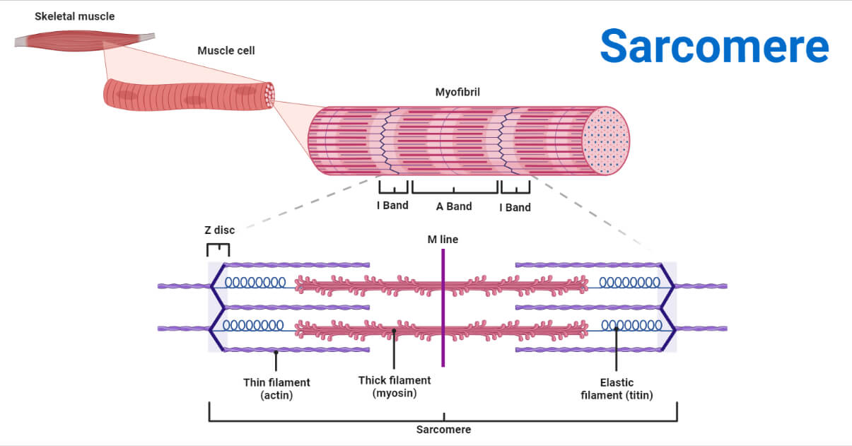
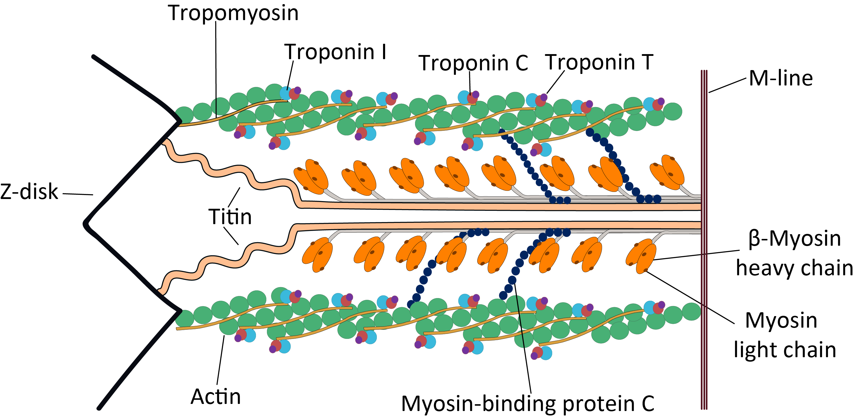
Sarcomere Drawing Labeled - Learn vocabulary, terms, and more with flashcards, games, and other study tools. Label the parts of the sarcomere. A z disc forms the boundary of the sarcomere on. The sarcomere fundamentally consists of two main myofilaments: In addition to myosin and actin, several other proteins, such as tropomyosin,. Web muscles work on a macro level, starting with tendons that attach muscles to bones. Web a sarcomere (greek σάρξ sarx flesh, μέρος meros part) is the smallest functional unit of striated muscle tissue. Web start studying sarcomere labeled diagram. A sarcomere is composed of two main protein filaments (thin actin and thick myosin filaments) which are the active structures responsible for muscular contraction. It is made up of multiple myosin and actin filaments oriented in parallel.
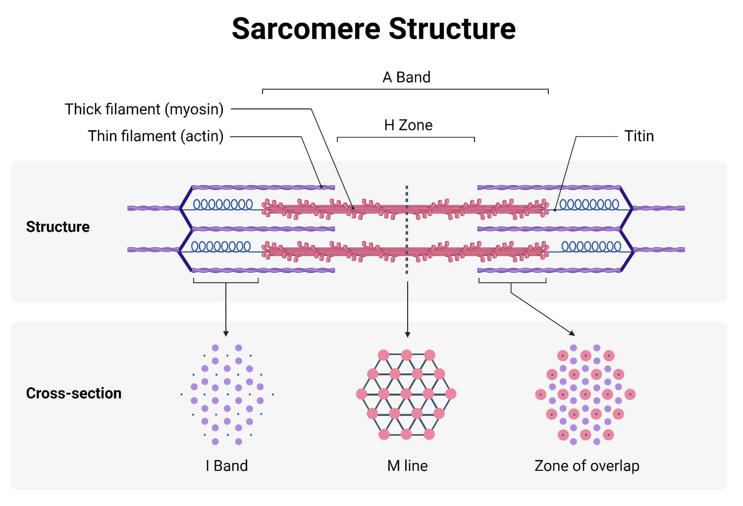
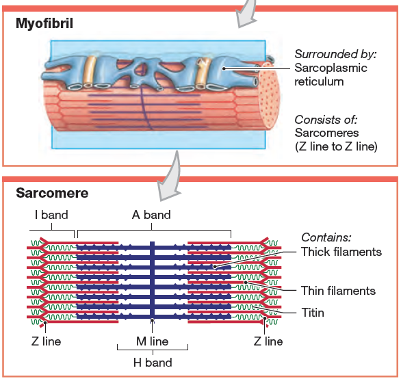
A sarcomere is a highly organized structure made up of thick and thin protein filaments; Learn vocabulary, terms, and more with flashcards, games, and other study tools. These layers cover muscle subunits, individual muscle cells, and myofibrils respectively. (b) a conceptual diagram representing the connectivity of molecules within a sarcomere. Web the sarcomere is the functional unit of a skeletal muscle cell. Sarcomeres are the basic contractile units of striated muscle cells. Web the fundamental repeat unit within muscle that is responsible for contraction is the sarcomere. The right side (pink color) of the sarcomere reflects a half sarcomere in. This is a distinguishing unit in some types of muscle tissue. It is represented as a thin, dark line in.
The neuromuscular junction another specialization of the skeletal muscle is the site where a motor neuron’s terminal meets the muscle fiber—called the neuromuscular junction (nmj). Web the region labeled with a z is called the a band. A person standing between two bookcases (z bands) pulls them in via. Sarcomeres are the basic contractile units of striated muscle cells. Web learn how to draw a labeled diagram of the structure of sarcomere, the basic unit of muscle contraction, with this easy and clear tutorial video. They first observed that the dynamic changes that were taking place were always happening in the same spots, or zones. Web a sarcomere is a microscopic segment repeating in a myofibril. These layers cover muscle subunits, individual muscle cells, and myofibrils respectively. Anatomical is said to be the term of microanatomy. Web a sarcomere is the basic contractile unit of a myocyte (muscle fibre).
Definition, Structure, Diagram, and Functions
The structure of the sarcomere is traditionally. Web the sarcomere is the functional unit of a skeletal muscle cell. Web dodge durango wiring diagram. Having a clear visual representation of a sarcomere can greatly aid in understanding its complex structure and functions. A sarcomere is composed of two main protein filaments (thin actin and thick myosin filaments) which are the.

Schematic of structure. are the functional units
The sarcomere is the basic contractile unit for both striated and cardiac muscle and is made up of a complex mesh of thick filaments, thin filaments, and a giant. Web the contractile unit of skeletal muscles. Web the fundamental repeat unit within muscle that is responsible for contraction is the sarcomere. The a band encompasses the h zone, but it.
Learn vocabulary, terms, and more with flashcards, games, and other study tools. Due to the striated nature of both skeletal muscle and cardiac muscle is observed by microscope slides. The sarcomere is the basic contractile unit for both striated and cardiac muscle and is made up of a complex mesh of thick filaments, thin filaments, and a giant. Web the.
Definition, Structure, Diagram, and Functions
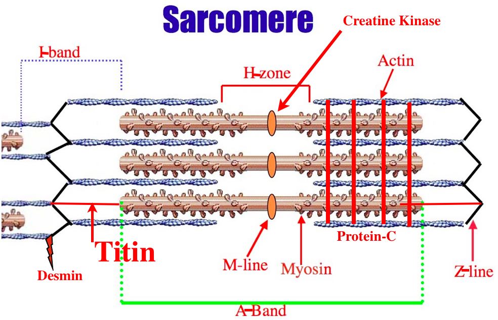
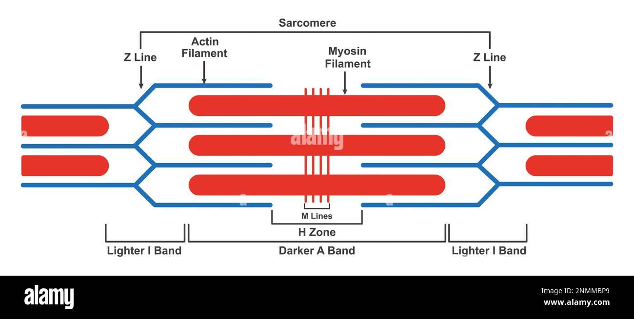
Learn vocabulary, terms, and more with flashcards, games, and other study tools. The actin and myosin filaments overlap in certain places creating several bands and zones. Thick filaments called myosin and thin filaments called actin. This is a distinguishing unit in some types of muscle tissue. Within muscles, there are layers of connective tissue called the epimysium, perimysium, and endomysium.
structure, illustration Stock Photo Alamy
A z disc forms the boundary of the sarcomere on. Label the parts of the brain. The sarcomere is the basic unit function with muscle fiber cells. The sarcomere fundamentally consists of two main myofilaments: Due to the striated nature of both skeletal muscle and cardiac muscle is observed by microscope slides.

Contracted Diagram
It is represented as a thin, dark line in. The a band encompasses the h zone, but it also contains regions around its outer edges where actin and myosin overlap, which makes these regions appear slightly darker. Definition, structure, diagram, and functions. The length of a single sarcomere is measured as the distance between two z lines, which on this.
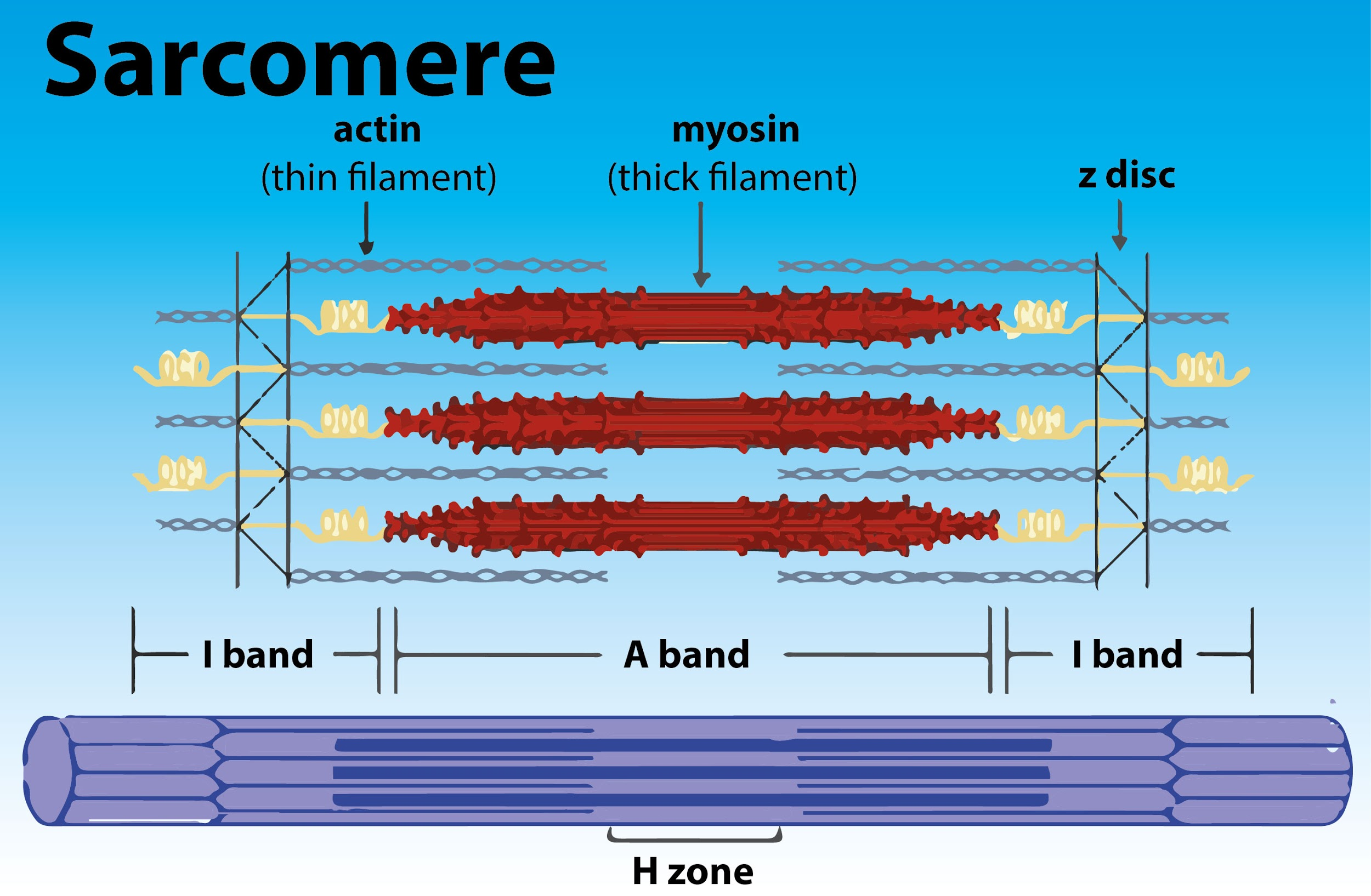
Diagram Labeled
The sarcomere is the basic contractile unit for both striated and cardiac muscle and is made up of a complex mesh of thick filaments, thin filaments, and a giant. Web the contractile unit of skeletal muscles. The right side (pink color) of the sarcomere reflects a half sarcomere in. Web a sarcomere is a microscopic segment repeating in a myofibril..
[Solved] 12. Draw and label the parts of a Course Hero
Web the sarcomere is the functional unit of a skeletal muscle cell. Web a sarcomere is a microscopic segment repeating in a myofibril. Web the figure depicts the structure of a sarcomere. Thick filaments called myosin and thin filaments called actin. A sarcomere is composed of two main protein filaments (thin actin and thick myosin filaments) which are the active.
FileCardiac structure.png Wikimedia Commons
Web muscles work on a macro level, starting with tendons that attach muscles to bones. Web actin and the z discs are shown in red. Sarcomeres are the basic units of muscle contraction and are responsible for the muscle’s ability to generate force. These layers cover muscle subunits, individual muscle cells, and myofibrils respectively. Learn vocabulary, terms, and more with.
Definition, Structure, & Sliding Filament Theory
Thick filaments called myosin and thin filaments called actin. Label the parts of the brain. Web the fundamental repeat unit within muscle that is responsible for contraction is the sarcomere. It is represented as a thin, dark line in. (b) a conceptual diagram representing the connectivity of molecules within a sarcomere.
The Sarcomere Fundamentally Consists Of Two Main Myofilaments:
Web the region labeled with a z is called the a band. The a band encompasses the h zone, but it also contains regions around its outer edges where actin and myosin overlap, which makes these regions appear slightly darker. A person standing between two bookcases (z bands) pulls them in via. Web the contractile unit of skeletal muscles.
Definition, Structure, Diagram, And Functions.
Web the fundamental repeat unit within muscle that is responsible for contraction is the sarcomere. Learn vocabulary, terms, and more with flashcards, games, and other study tools. Due to the striated nature of both skeletal muscle and cardiac muscle is observed by microscope slides. In addition to myosin and actin, several other proteins, such as tropomyosin,.
Learn Vocabulary, Terms, And More With Flashcards, Games, And Other Study Tools.
Web the sarcomere is the functional unit of a skeletal muscle cell. They first observed that the dynamic changes that were taking place were always happening in the same spots, or zones. Thick filaments called myosin and thin filaments called actin. A z disc forms the boundary of the sarcomere on.
Web The Figure Depicts The Structure Of A Sarcomere.
The widely accepted theory describing muscular contraction is called the sliding filament theory, which proposes that. Within muscles, there are layers of connective tissue called the epimysium, perimysium, and endomysium. Web a sarcomere (greek σάρξ sarx flesh, μέρος meros part) is the smallest functional unit of striated muscle tissue. The sarcomere is the basic unit function with muscle fiber cells.