Serous Membrane Drawing
Serous Membrane Drawing - The pleural cavity reduces friction. Web the pleural fluid helps lubricate the membranes lining the pleural cavity and lungs when breathing in and out. A serous membrane consists of a single layer of flattened mesothelial cells applied to the Web the serous membrane. Resident progenitors, not exogenous migratory cells, generate the majority of visceral mesothelium in organogenesis. Web membranes of the anterior (ventral) body cavity. Serous membranes consist of a single layer of epithelium, named mesothelium, attached to a supporting layer of connective tissue, with a small layer in between, the basal membrane (fig 1). Mesothelium is composed of specialized mesothelial cells which produce serous fluid, a thin, watery. Both the parietal and visceral serosa secrete the thin, slippery serous fluid located within the serous cavities. Body cavities and serous membranes.
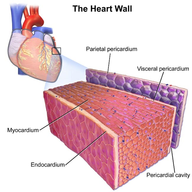
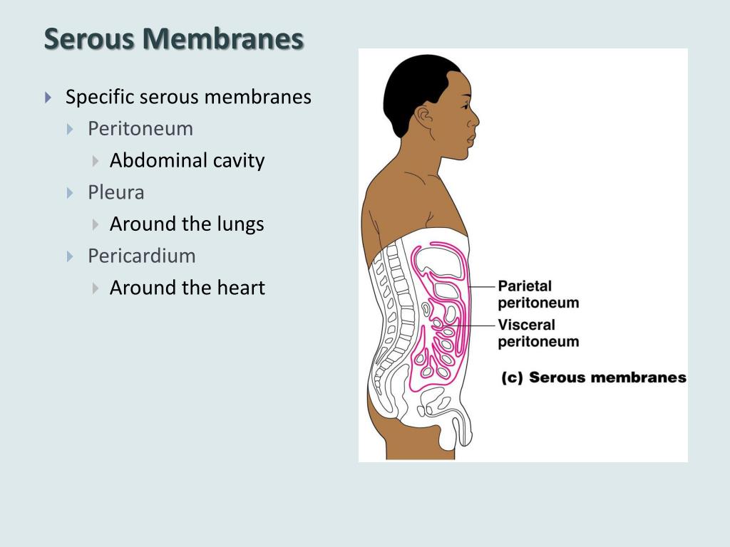
Both the parietal and visceral serosa secrete the thin, slippery serous fluid located within the serous cavities. These membranes line the coelomic cavities of the body, and they cover the organs located within those cavities. Web serous membranes are identified according locations. Web the peritoneum is the serous membrane that forms the lining of the abdominal cavity or the coelom. Any serous membrane will always have two parts: Web mesothelium is a simple squamous epithelium that forms the epithelial layer of serous membranes which line body cavities and internal organs.serous membranes, and therefore mesothelium, line the pericardial, pleural and peritoneal cavities. The two pleura that cover the lungs and the pericardium that covers the heart. One is covering the outermost surface of the organ, and is call the visceral layer. Serous membranes consist of a single layer of epithelium, named mesothelium, attached to a supporting layer of connective tissue, with a small layer in between, the basal membrane (fig 1). We shall now consider the structures of the pleurae in more detail.
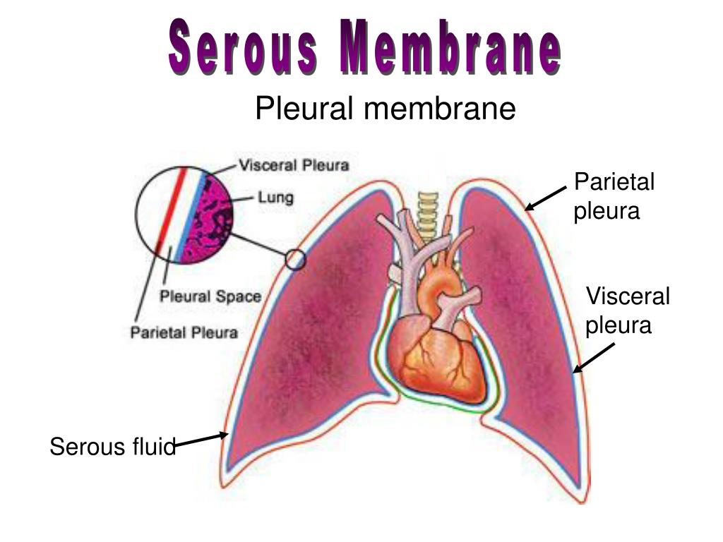
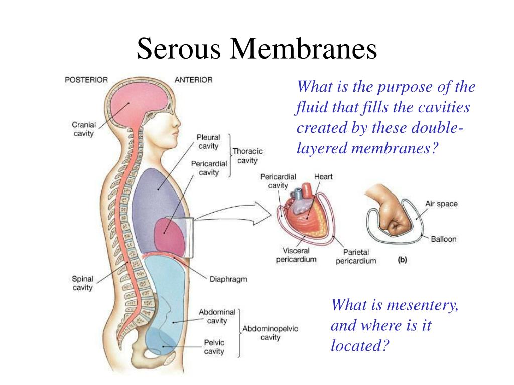
The two pleura that cover the lungs and the pericardium that covers the heart. Web the pleural fluid helps lubricate the membranes lining the pleural cavity and lungs when breathing in and out. Each pleural cavity is lined by a serous membrane called the pleura. Both the parietal and visceral serosa secrete the thin, slippery serous fluid located within the serous cavities. Web serous membranes are identified according locations. The pleural fluid is located in the pleural cavity, also known as the pleural space, which is the potential space between the lungs and chest wall shown in yellow. Three serous membranes line the thoracic cavity; Web mesothelium is a simple squamous epithelium that forms the epithelial layer of serous membranes which line body cavities and internal organs.serous membranes, and therefore mesothelium, line the pericardial, pleural and peritoneal cavities. Web the serosal mesothelium is a major source of smooth muscle cells of the gut vasculature. Web the serosa, also known as the serous membrane, is a single layer of simple squamous epithelium called mesothelium.it is supported by a thin underlying layer of loose connective tissue, abundant in blood vessels, lymphatic vessels, nerves and adipose tissue.it lines closed body cavities including the pericardial, peritoneal and pleural.

serous membranes of thoracic Cavity (part 1) Diagram Quizlet
Web the pleural fluid helps lubricate the membranes lining the pleural cavity and lungs when breathing in and out. The two pleura that cover the lungs and the pericardium that covers the heart. This also explains the name 'serous membrane'. It is composed of a layer of mesothelial tissue, supported by a thin layer of connective tissue. We shall now.
Serous Membrane Definition, Function and Structure Biology Dictionary
Winters, ni, williams, am, bader, dm. Resident progenitors, not exogenous migratory cells, generate the majority of visceral mesothelium in organogenesis. The pleural fluid is located in the pleural cavity, also known as the pleural space, which is the potential space between the lungs and chest wall shown in yellow. Any serous membrane will always have two parts: In anatomy, the.

Chapter 1I Serous Membranes YouTube
The thin membrane is made up of mesothelium tissue which originates from the mesoderm. Three serous membranes line the thoracic cavity; Web serous membrane cavities — are lined by serous membrane — are normally empty (except for microscopic cells and a film of fluid) — function to preclude adhesions among organs, thereby allowing organs to move freely relative to one.

Anatomy Body Cavities & Serous Membranes YouTube
One is covering the outermost surface of the organ, and is call the visceral layer. Three serous membranes line the thoracic cavity; Body cavities and serous membranes. Web the pleural fluid helps lubricate the membranes lining the pleural cavity and lungs when breathing in and out. These two parts are continuous with each other at the hilum of each lung.
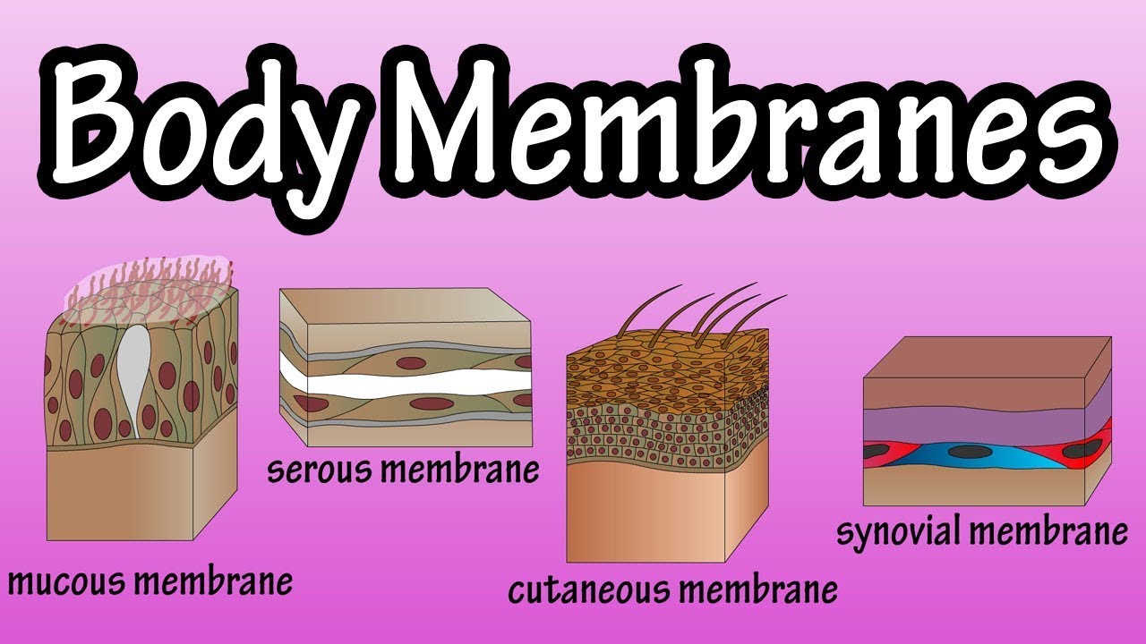
Body Membranes Types Of Membranes In The Body Serous Membranes
Resident progenitors, not exogenous migratory cells, generate the majority of visceral mesothelium in organogenesis. Serous membranes consist of a single layer of epithelium, named mesothelium, attached to a supporting layer of connective tissue, with a small layer in between, the basal membrane (fig 1). Three serous membranes line the thoracic cavity; Web draw it video tutorial: Web the serous membrane,.
PPT The Human Body PowerPoint Presentation ID640744
Histology of a serous membrane. Web the ventral body cavities are enclosed inside serous membranes ( serosa) that line the cavities and the organs inside them and create fluids that lubricate cushion the cavities to protect the organs prevent friction during movement. Any serous membrane will always have two parts: Parietal serosa line the body cavities and visceral serosa line.

Serous Membrane, Vintage Illustration Stock Vector Illustration of
The two pleura that cover the lungs and the pericardium that covers the heart. Any serous membrane will always have two parts: The pleural fluid is located in the pleural cavity, also known as the pleural space, which is the potential space between the lungs and chest wall shown in yellow. Body cavities and serous membranes. Each pleural cavity is.
PPT Skin and Body Membranes PowerPoint Presentation, free download
The two pleura that cover the lungs and the pericardium that covers the heart. The serous membrane allows for frictionless movement in a number of vital organs. Each pleural cavity is lined by a serous membrane called the pleura. Three serous membranes line the thoracic cavity; Both the parietal and visceral serosa secrete the thin, slippery serous fluid located within.
Body Cavities and Membranes SCIENTIST CINDY
It is composed of a layer of mesothelial tissue, supported by a thin layer of connective tissue. Web the pleural fluid helps lubricate the membranes lining the pleural cavity and lungs when breathing in and out. In histology this layer is called serosa after serous membrane. Web the serous membrane in the drawing above is composed of epithelium which is.
PPT The Tissue Level of Organization PowerPoint Presentation, free
The serous membrane allows for frictionless movement in a number of vital organs. The two pleura that cover the lungs and the pericardium that covers the heart. The two pleura that cover the lungs and the pericardium that covers the heart. Web serous membranes are identified according locations. Web the serosa, also known as the serous membrane, is a single.
The Peritoneum Provides Support And Protection For The Abdominal Organs, And Is The.
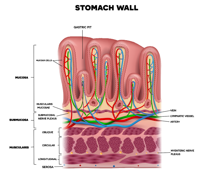
Three serous membranes line the thoracic cavity; Histologically, this can be seen as a layer on the outside of the gut. A fourth, the peritoneum, is the serous membrane in the abdominal cavity that covers abdominal organs and forms double sheets of mesenteries that suspend many of the. Web the serous membrane in the drawing above is composed of epithelium which is always simple squamous epithelium attached to a thin layer of connective tissue, which is always areolar connective tissue.
The Serous Membrane Allows For Frictionless Movement In A Number Of Vital Organs.
In anatomy, the serous membrane (or serosa) is a smooth membrane that consists of a thin connective tissue layer and a thin layer of cells that secrete serous fluid. Body cavities and serous membranes. It is composed of a layer of mesothelial tissue, supported by a thin layer of connective tissue. Web serous membranes are identified according locations.
Web The Pleural Fluid Helps Lubricate The Membranes Lining The Pleural Cavity And Lungs When Breathing In And Out.
Mesothelium is composed of specialized mesothelial cells which produce serous fluid, a thin, watery. Each pleural cavity is lined by a serous membrane called the pleura. The pleural fluid is located in the pleural cavity, also known as the pleural space, which is the potential space between the lungs and chest wall shown in yellow. Web anatomy physiology of body cavities, serous membranes, and their contents explained, drawn, defined.
Three Serous Membranes Line The Thoracic Cavity;
The two pleura that cover the lungs and the pericardium that covers the heart. Web membranes of the anterior (ventral) body cavity. A serous membrane (also referred to as serosa) is an epithelial membrane composed of mesodermally derived epithelium called the mesothelium that is supported by connective tissue. In histology this layer is called serosa after serous membrane.