Simple Columnar Epithelium Drawing
Simple Columnar Epithelium Drawing - The simple columnar epithelium has a wide variety of functions,. Instead of being smooth, the inside of the intestine is folded and covered by millions of tiny projections called villi. Web the inner surface of the intestinal wall is made of simple columnar epithelium (sce). Web drawing histological diagram of simple columnar epithelia.useful for all medical students.drawn by using h & e pencils.explanation on epithelia while drawing. On the image, you can see one fold on the right and parts of several villi sticking up from the surface of the fold. Epithelial tissue is often classified according to numbers of layers of cells present, and by the shape of the cells. Web simple columnar epithelium is found mainly in the digestive system comprising the endothelial lining of the stomach, small intestine, large intestine, and gall bladder. These columnar epithelium consist of tall columnar cells. Read more about simple collumnar epithelium 10x; Trachea and most of the upper respiratory tract (ciliated cells) function:
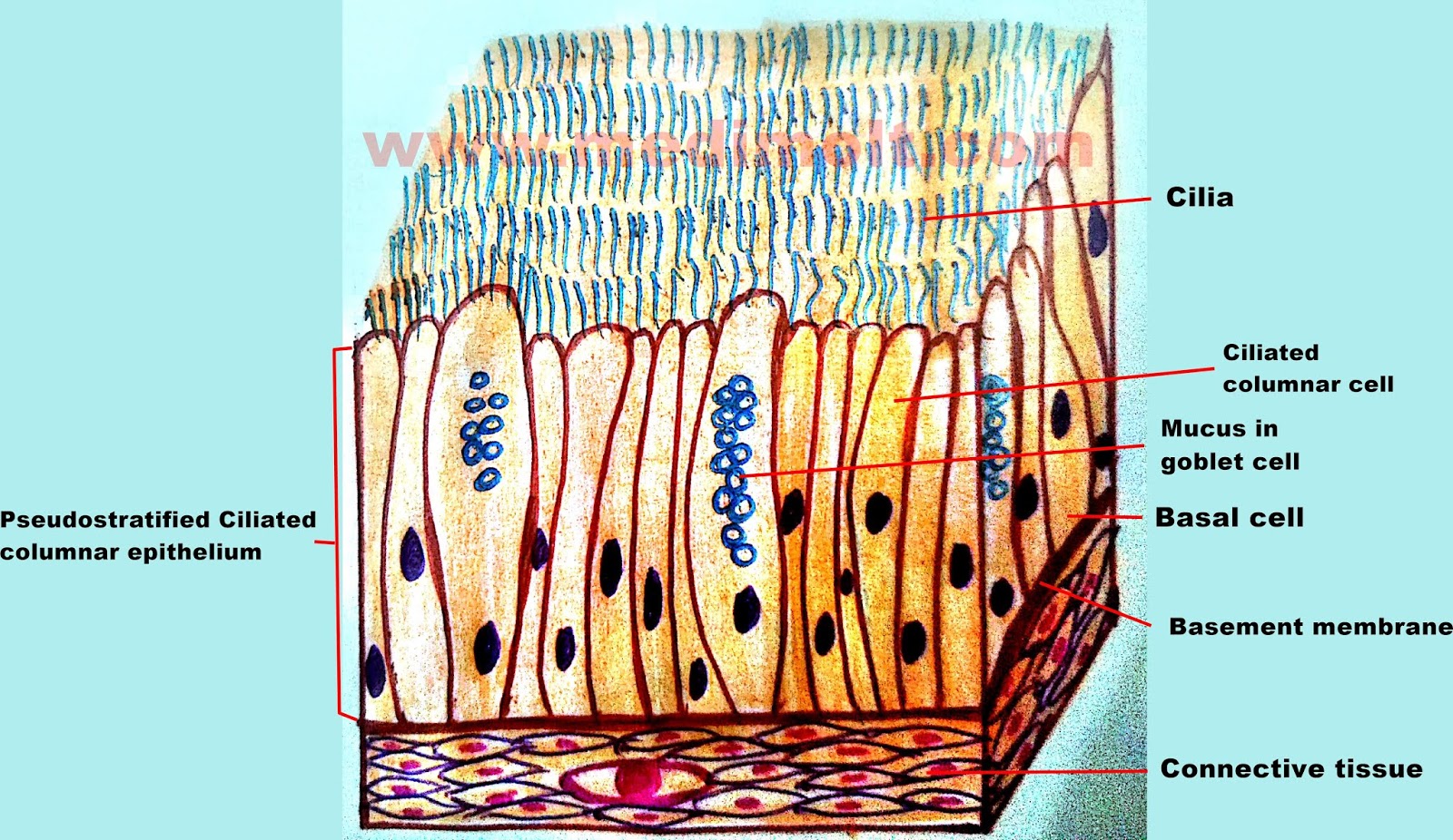
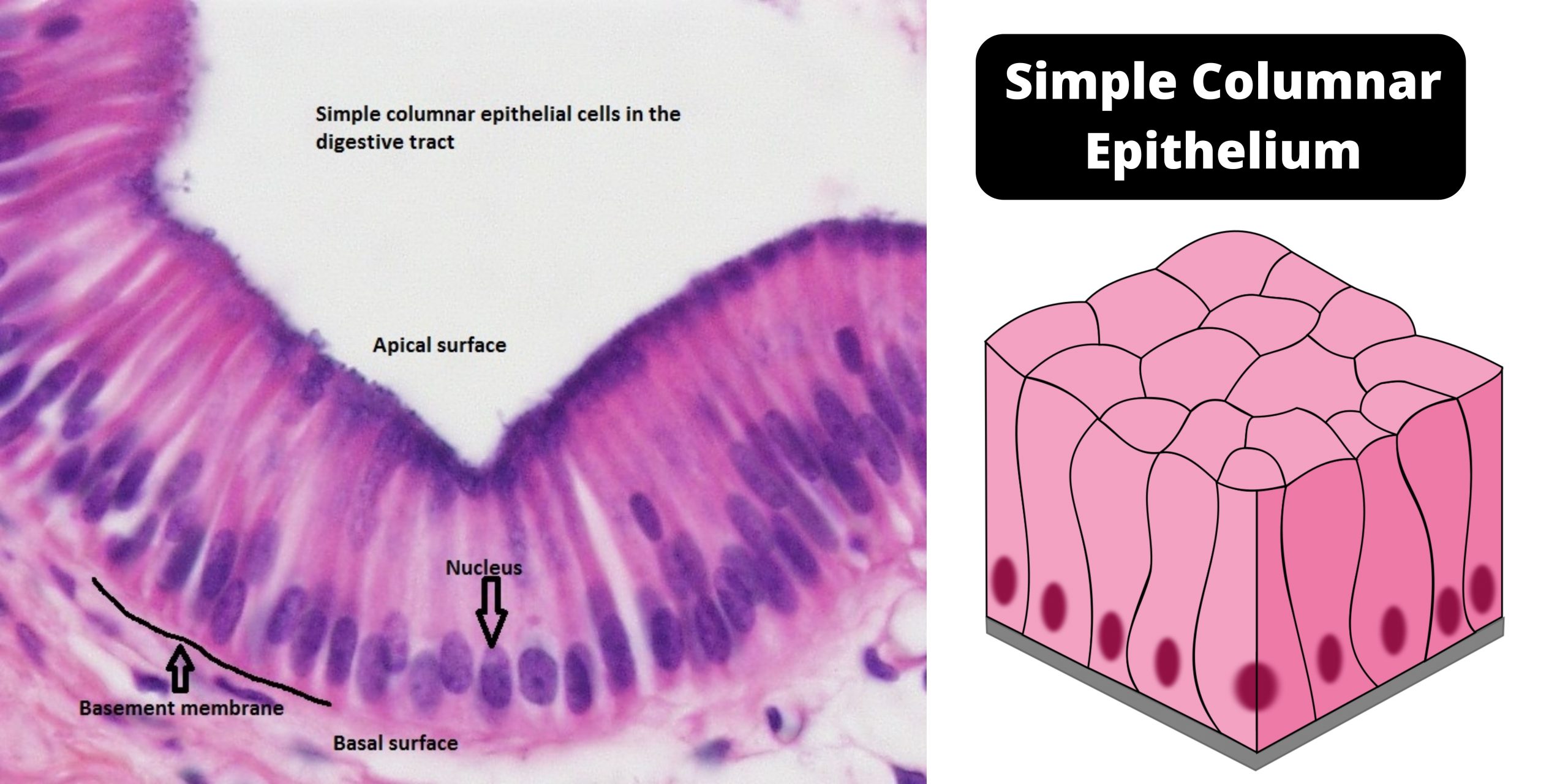
Epithelial tissue is often classified according to numbers of layers of cells present, and by the shape of the cells. The basement membrane is a thin but strong, acellular layer which lies between the epithelium and the adjacent connective tissue. In humans, simple columnar epithelium lines most organs of the digestive tract including the stomach, and intestines. Every cell attaches to the basement membrane. Ciliated columnar epithelium is composed of simple columnar epithelial cells with cilia on their apical surfaces. Where the section was cut orthogonal to the surface of the villi, a single row of cells are seen. These epithelial cells are found in the lining of the fallopian tubes where the assist in the passage of the egg. Web sweat glands, salivary glands, mammary glands, adrenal glands, and pituitary glands are examples of glands made of epithelial tissue. Trachea and most of the upper respiratory tract (ciliated cells) function: Again, if you want to see these simple columnar epithelium as the verticle or transverse section, you will find them rectangular.
And so notice that these simple cuboidal epithelial tissue cells are forming a ring here because they're forming the tubules of the kidney. Web schematic drawing of the simple columnar epithelium. Like the cuboidal epithelia, this epithelium is active in the absorption and secretion of molecules. The cells that make up this epithelium are taller than they are large and their nuclei, positioned in the lower third of the cytoplasm, are elongated. Web read more about simple columnar epithelium 20x; The simple columnar epithelium has a wide variety of functions,. Web the inner surface of the intestinal wall is made of simple columnar epithelium (sce). Is a single layer of cells that are taller than they are wide. Web but when we draw a sketch of the same micrograph, it makes it much more easier to see the simple cuboidal epithelial tissue cells. On the image, you can see one fold on the right and parts of several villi sticking up from the surface of the fold.
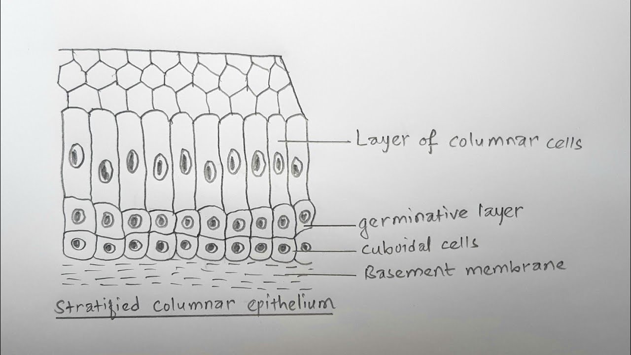
How to draw stratified columnar epithelium easy way YouTube
Web read more about simple columnar epithelium 20x; Web simple columnar epithelium is found mainly in the digestive system comprising the endothelial lining of the stomach, small intestine, large intestine, and gall bladder. Every cell attaches to the basement membrane. Web simple columnar epithelium forms a majority of the digestive tract and some parts of the female reproductive tract. In.

Simple Columnar Epithelium (LM) Stock Image C022/2221 Science
These columnar epithelium consist of tall columnar cells. The cells of this epithelium are arranged in a neat row with the nuclei at the same level, near the basal end. Simple columnar epithelium consists of a single layer of cells that are taller than they are wide, with an oval nucleus usually located towards the basal region of the cell..

Simple columnar epithelium Diagram Quizlet
Web drawing histological diagram of simple columnar epithelia.useful for all medical students.drawn by using h & e pencils.explanation on epithelia while drawing. In contrast, the ciliated columnar epithelium aids the transport or movement of molecules and cells from one place to. Ciliated columnar epithelium is composed of simple columnar epithelial cells with cilia on their apical surfaces. Web schematic drawing.
Ciliated Simple Columnar Epithelium Labeled Jajae Studio
Instead of being smooth, the inside of the intestine is folded and covered by millions of tiny projections called villi. These cells are placed side by s. Where the section was cut orthogonal to the surface of the villi, a single row of cells are seen. These columnar epithelium consist of tall columnar cells. Web drawing histological diagram of simple.
Simple columnar epithelium definition, structure, functions, examples
Simple columnar epithelium consists of a single layer of cells that are taller than they are wide, with an oval nucleus usually located towards the basal region of the cell. In humans, simple columnar epithelium lines most organs of the digestive tract including the stomach, and intestines. It also secretes mucus, which helps to lubricate, moisten, and protect the surface..

Simple columnar epithelium, light micrograph Stock Image C052/8835
It also secretes mucus, which helps to lubricate, moisten, and protect the surface. The basement membrane is a thin but strong, acellular layer which lies between the epithelium and the adjacent connective tissue. In the small intestine, it facilitates the absorption of nutrients. Web read more about simple columnar epithelium 20x; Web simple columnar epithelium forms a majority of the.

Simple columnar epithelium Pseudostratified columnar epithelium Simple
Ciliated columnar epithelium is composed of simple columnar epithelial cells with cilia on their apical surfaces. The dark line underneath the brush border is the terminal web in which the microvilli are anchored. Web but when we draw a sketch of the same micrograph, it makes it much more easier to see the simple cuboidal epithelial tissue cells. The cells.

Simple Columnar Epithelium 40x Histology
Web simple columnar epithelium is found mainly in the digestive system comprising the endothelial lining of the stomach, small intestine, large intestine, and gall bladder. These cells are placed side by s. Digitally annotated micrograph of the columnar epithelium. Web read more about simple columnar epithelium 20x; In the small intestine, it facilitates the absorption of nutrients.

Simple columnar epithelium Diagram Quizlet
Read more about simple collumnar epithelium 10x; Web simple columnar epithelia are tissues made of a single layer of long epithelial cells that are often seen in regions where absorption and secretion are important features. These columnar epithelium consist of tall columnar cells. It is sometimes referred to as the “basal lamina”. These epithelial cells are found in the lining.

Simple Columnar Epithelium Description Single layer of elongated
Web structures and types of simple epithelia. Read more about simple collumnar epithelium 10x; Web read more about simple columnar epithelium 20x; Every cell attaches to the basement membrane. Please include total magnification in the image key.
And So We Can Say That Simple Columnar Epithelium Consists Of Just One.
It also secretes certain enzymes. Web drawing histological diagram of simple columnar epithelia.useful for all medical students.drawn by using h & e pencils.explanation on epithelia while drawing. Instead of being smooth, the inside of the intestine is folded and covered by millions of tiny projections called villi. And so notice that these simple cuboidal epithelial tissue cells are forming a ring here because they're forming the tubules of the kidney.
It Also Secretes Mucus, Which Helps To Lubricate, Moisten, And Protect The Surface.
Please include total magnification in the image key. A simple epithelium is only one layer of cells thick. The basement membrane is a thin but strong, acellular layer which lies between the epithelium and the adjacent connective tissue. Web on a concluding note, simple columnar epithelium has two primary functions of absorption and secretion.
Allows Absorbtion, Secretes Mucous And Enzymes.
Web schematic drawing of the simple columnar epithelium. It is sometimes referred to as the “basal lamina”. Trachea and most of the upper respiratory tract (ciliated cells) function: Web simple columnar epithelium forms a majority of the digestive tract and some parts of the female reproductive tract.
Where The Section Was Cut Orthogonal To The Surface Of The Villi, A Single Row Of Cells Are Seen.
The dark line underneath the brush border is the terminal web in which the microvilli are anchored. Web but when we draw a sketch of the same micrograph, it makes it much more easier to see the simple cuboidal epithelial tissue cells. On the image, you can see one fold on the right and parts of several villi sticking up from the surface of the fold. The simple columnar epithelium has a wide variety of functions,.