Spinal Cord Drawing Easy
Spinal Cord Drawing Easy - Determine the motor or sensory loss that would be expected from interruption of each of the three tracts. They are analogous with the cranial meninges. It extends from the external margin of the foramen magnum as a continuation of the medulla oblongata, down to the l2 vertebral level, and is entirely housed in the spinal meningeal layers. Web your spinal cord is the long, cylindrical structure that connects your brain and lower back. Cervical enlargement, lumbosacral enlargement, medullary cone, spinal part of filum terminale, cauda equina, spinal nerves. Describe the type of information carried by each of the three tracts. A cutaway view of the rachidian nerve highlights its structure with from the outside. Draw the location of each of these tracts within the spinal cord. I have a spinal cord flattening in my cervical spine, a leaked disc, a torn rotator cuff that won’t heal and a dislocating shoulder which won’t allow it to heal. As the spinal cord transitions through the cervical and.
It is a relatively small bundle of tissue (weighing 35g and just about 1cm in diameter) but is crucial in facilitating our daily activities. Determine the location of a spinal cord lesion from a given motor and/or sensory loss. Web spinal cord, drawing the spinal cord. During development, there’s a disproportion between spinal cord growth and vertebral column growth. I have a spinal cord flattening in my cervical spine, a leaked disc, a torn rotator cuff that won’t heal and a dislocating shoulder which won’t allow it to heal. This part of the spine is known as the lumbar region. Texture the bone with additional curved lines. Web anatomy of the spinal cord. Describe the type of information carried by each of the three tracts. Web by the end of this session, learners will be able to:
During development, there’s a disproportion between spinal cord growth and vertebral column growth. It contains tissues, fluids and nerve cells. The central nervous system, consisting of the brain and spinal cord, and the peripheral nervous system, made up of nerves and ganglia. I am sick of being sick. Web spine pelvic floor print, physical therapy art, pelvic health art, abstract pelvis line art, chropractic poster physical therapy. A cutaway view of the rachidian nerve highlights its structure with from the outside. Many more tests need to be ran. Autopsy, surgery educational cards, drawing, potion, vial artwork, horror prop, apothecary. Cervical enlargement, lumbosacral enlargement, medullary cone, spinal part of filum terminale, cauda equina, spinal nerves. Describe the type of information carried by each of the three tracts.

Pin on sketch art
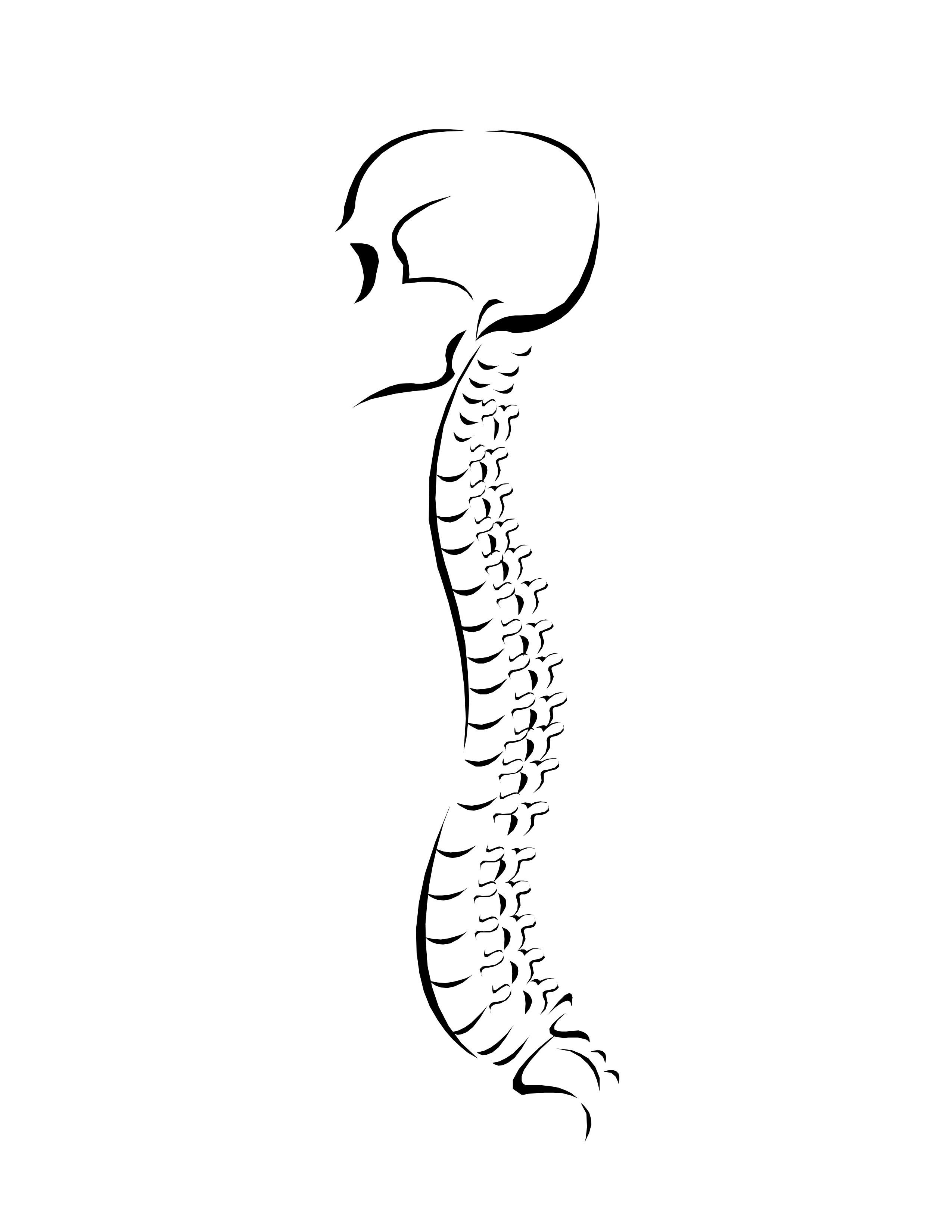
Easy way to draw human backbone skeleton diagram. It contains tissues, fluids and nerve cells. The spinal cord is part of the central nervous system (cns). The spinal cord is a cylinder that is roughly 45 cm long and 1 cm wide. A cutaway view of the rachidian nerve highlights its structure with from the outside.
Spine Drawing Easy For Kids Spine Drawing Simple Getdrawings Figure
Easy way to draw human backbone skeleton diagram. Web spine pelvic floor print, physical therapy art, pelvic health art, abstract pelvis line art, chropractic poster physical therapy. Web the spinal cord is part of the central nervous system and consists of a tightly packed column of nerve tissue that extends downwards from the brainstem through the central column of the.
Spinal Cord Drawing at GetDrawings Free download
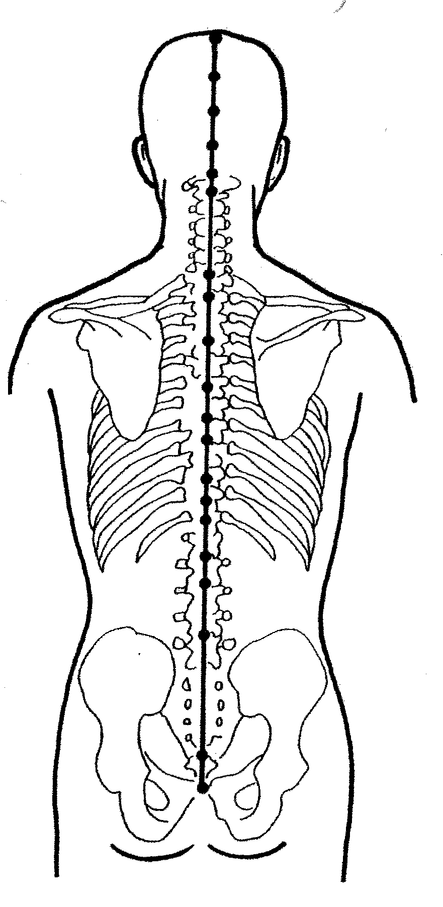
Also shown are the spinal cord, vertebra (back bone), conus medullaris (the end of the spinal cord), cauda equina (the. Web your spinal cord is the long, cylindrical structure that connects your brain and lower back. Information travels in two directions: It is situated inside the vertebral canal of the vertebral column. I have a spinal cord flattening in my.

Which of the following is responsible for protection of Spinal Cord
Recognize the three major tracts of the spinal cord: Web how to draw human spine drawing step by step easy / spinal cord diagram / vertebral column drawing / spine drawing / ceragem spine chart / read ki haddi ka c. I have a spinal cord flattening in my cervical spine, a leaked disc, a torn rotator cuff that won’t.

Spinal Cord USMLE Strike
My insurance is 500 a month and visits to specialists are 40$. Representation in 3/4 front view of the stucture of the spinal cord, and rachidian nerves. They are analogous with the cranial meninges. The nervous system is divided into two main parts: Web how to draw human spine anatomy step by step very easy.

spine Anatomy Sketches, Anatomy Drawing, Anatomy Art, Human Anatomy
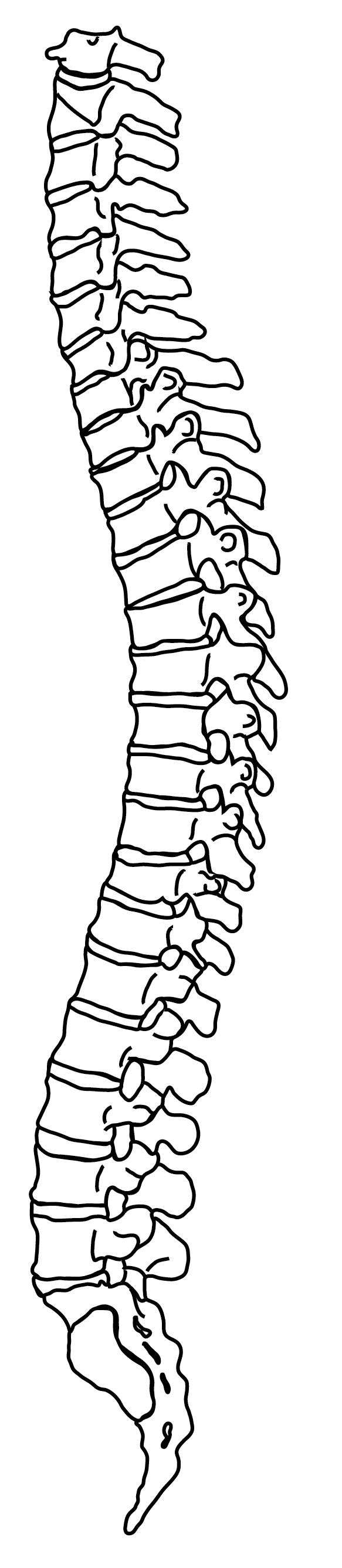
Web the spinal cord can be equally divided along the midline dorsoventral axis by drawing a line through the depression known as the dorsal and ventral median sulci.on each half of the spinal cord, a ventrolateral and dorsolateral sulcus is appreciated at the sites from which the ventral and dorsal nerve roots leave and enter the spinal cord,. Then, begin.

How to Draw Structure Of The Spinal Cord Diagram Easy And Step by Step
Web how to draw human spine drawing step by step easy / spinal cord diagram / vertebral column drawing / spine drawing / ceragem spine chart / read ki haddi ka c. Then, begin drawing the vertebra. Texture the bone with additional curved lines. A bony column of vertebrae surrounds and protects your spinal cord. Web by the end of.
Simple Spine Drawing at Explore collection of
Web your spinal cord is the long, cylindrical structure that connects your brain and lower back. Subscribe to my channel to get more drawing. Web old engraved illustration of human skeletons. Easy way to draw human backbone skeleton diagram. From the spinal cord se dã©tachent on each side rachidian nerves constituted of a ganglion, a posterior and an anterior root.

How To Draw Spinal Cord Step By Step Easily YouTube
Distally, the meninges form a strand of fibrous tissue, the filum. Representation in 3/4 front view of the stucture of the spinal cord, and rachidian nerves. The nervous system is divided into two main parts: It contains tissues, fluids and nerve cells. Web the spinal cord can be equally divided along the midline dorsoventral axis by drawing a line through.
![[Biology Class 10] Spinal Cord Structure, Function, Diagram](https://d77da31580fbc8944c00-52b01ccbcfe56047120eec75d9cb2cbd.ssl.cf6.rackcdn.com/c02a0a4e-2c36-414c-9461-ddc6860c952d/spinal-cord---teachoo.jpg)
[Biology Class 10] Spinal Cord Structure, Function, Diagram
My insurance is 500 a month and visits to specialists are 40$. They contain cerebrospinal fluid, acting to support and protect the spinal cord. Web how to draw human spine anatomy step by step very easy. The central nervous system, consisting of the brain and spinal cord, and the peripheral nervous system, made up of nerves and ganglia. I am.
Determine The Location Of A Spinal Cord Lesion From A Given Motor And/Or Sensory Loss.
My insurance is 500 a month and visits to specialists are 40$. Spinal cord drawing stock photos are available in a variety of sizes and formats to fit your needs. Many more tests need to be ran. Web by the end of this session, learners will be able to:
Web The Spinal Cord Can Be Equally Divided Along The Midline Dorsoventral Axis By Drawing A Line Through The Depression Known As The Dorsal And Ventral Median Sulci.on Each Half Of The Spinal Cord, A Ventrolateral And Dorsolateral Sulcus Is Appreciated At The Sites From Which The Ventral And Dorsal Nerve Roots Leave And Enter The Spinal Cord,.
They are analogous with the cranial meninges. From the periphery to the central nervous system via afferent neurons, and from the central nervous system to. Also shown are the spinal cord, vertebra (back bone), conus medullaris (the end of the spinal cord), cauda equina (the. Describe the type of information carried by each of the three tracts.
The Spinal Cord Is Part Of The Central Nervous System (Cns).
Web anatomy of the spinal cord. Representation in 3/4 front view of the stucture of the spinal cord, and rachidian nerves. Web draw the location of each of these tracts within the spinal cord. Web the spinal cord is part of the central nervous system and consists of a tightly packed column of nerve tissue that extends downwards from the brainstem through the central column of the spine.
They Contain Cerebrospinal Fluid, Acting To Support And Protect The Spinal Cord.
Web spinal cord, drawing the spinal cord. Autopsy, surgery educational cards, drawing, potion, vial artwork, horror prop, apothecary. Web how to draw spinal cord diagram easily and step by step tutorial by abhishek educare#abhishekeducarethanks for watching 🙏 Spinal cord functions include carrying signals between the brain.