Stratified Squamous Epithelium Drawing
Stratified Squamous Epithelium Drawing - The stratified epithelium is named by the shape of the most apical layer of cells, closest to the free space. Web 59,228 x 34,921 pixels 7.7 gb. The term “squamous” is derived from the comparison of the cells to the fish’s scales. Stratified squamous epithelium is the most common type of stratified. The keratinization, or lack thereof, of the apical surface domains of the cells. Click on to move to a specific region. O describe the structure of microvilli, cilia, and other apical specializations of epithelial cells. The top layer may be covered with dead cells containing keratin. Use the image slider below to learn how to use a microscope to identify and study nonkeratinized stratified squamous epithelium lining. Stratified squamous epithelia are tissues formed from multiple layers of cells resting on a basement membrane, with the superficial layer (s) consisting of squamous cells.
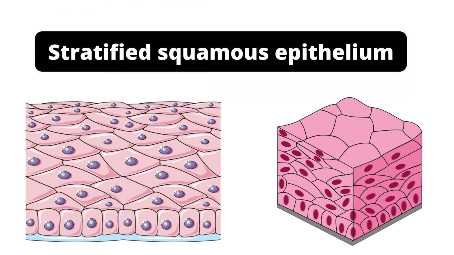
Web learn to draw stratified squamous keratinized epithelium histology diagram ( for mbbs and bds students) A stratified epithelium consists of several stacked layers of cells. Web the epidermis is composed of keratinized, stratified squamous epithelium. A typical example of stratified squamous keratinized epithelium is the epidermis. 19k views 2 years ago cell biology. The apical cells appear squamous, whereas the basal layer contains either columnar or cuboidal cells. Web stratified squamous epithelium is the most common type of stratified epithelium in the human body. Use the image slider below to learn more about the characteristics of stratified squamous epithelium. O classify morphological types of epithelia based on the number of cell layers, shape of apical cells, and presence of surface specializations. Medical school university of minnesota minneapolis, mn.
Web a stratified squamous epithelium consists of squamous (flattened) epithelial cells arranged in layers upon a basal membrane. Web here, i will provide both hand drawings and real microscope figures of stratified squamous epithelium (keratinized and nonkeratinized). This type of epithelium comprises the epidermis of the skin. They are flat and have an irregularly round shape. Web structure of the stratified squamous epithelium. Illustration of stratified squamous epithelium, showing surface layer, basal layer, basement membrane, and connective. Use the image slider below to learn more about the characteristics of stratified squamous epithelium. This video describes how to draw stratified squamous non keratinized epithelium histology diagram. Functions of the stratified squamous epithelium. Each slide is shown with additional information to its right.
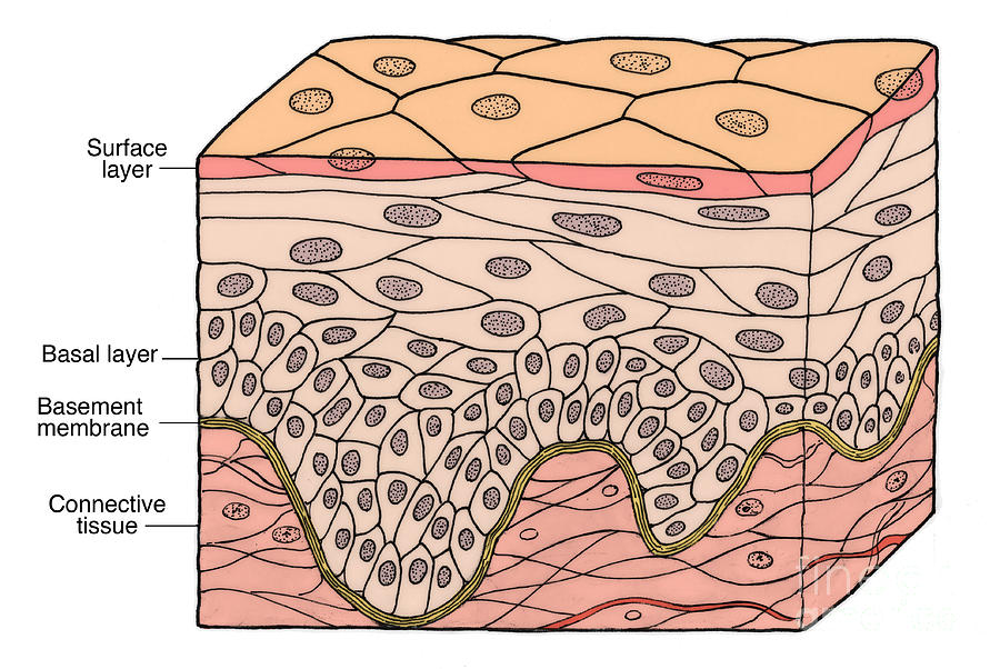
Illustration Of Stratified Squamous Photograph by Science Source Pixels
4.5k views 2 years ago. It does not have any blood vessels within it (i.e., it is avascular). The image can be changed using any combination of the following commands. Only one layer is in contact with the basement membrane; Location and examples of stratified squamous epithelium.

Nonkeratinized Stratified Squamous Epithelium Function And Structure
A typical example of stratified squamous keratinized epithelium is the epidermis. Illustration of stratified squamous epithelium, showing surface layer, basal layer, basement membrane, and connective. Functions of the stratified squamous epithelium. Only one layer is in contact with the basement membrane; Science source / science photo library.

Draw A Labelled Diagram Of Squamous Epithelial Tissue vrogue.co
51.0 mb (4.2 mb compressed) 5160 x 3455 pixels. Web stratified squamous epithelium is the most common type of stratified epithelium in the human body. Use the image slider below to learn more about the characteristics of stratified squamous epithelium. This type of epithelium comprises the epidermis of the skin. It does not have any blood vessels within it (i.e.,.
Epithelial tissues
Location and examples of stratified squamous epithelium. Use the image slider below to learn how to use a microscope to identify and study nonkeratinized stratified squamous epithelium lining. Simple columnar epithelium under a microscope. 3.8k views 3 years ago. The term “squamous” is derived from the comparison of the cells to the fish’s scales.

Stratified Squamous Epithelium in 2021 Stratified squamous epithelium
The apical cells appear squamous, whereas the basal layer contains either columnar or cuboidal cells. 51.0 mb (4.2 mb compressed) 5160 x 3455 pixels. Web simple squamous epithelium under microscope drawing. Web here, i will provide both hand drawings and real microscope figures of stratified squamous epithelium (keratinized and nonkeratinized). The other layers adhere to one another to maintain structural.

Stratified Squamous Keratinized Epithelium Diagram
Anatomy with amrutha & joseph. Web 59,228 x 34,921 pixels 7.7 gb. Location of simple columnar epithelium. Web when classifying a stratified epithelial sheet, the sheet is named for the shape of the cells in its most superficial layers. A typical example of stratified squamous keratinized epithelium is the epidermis.
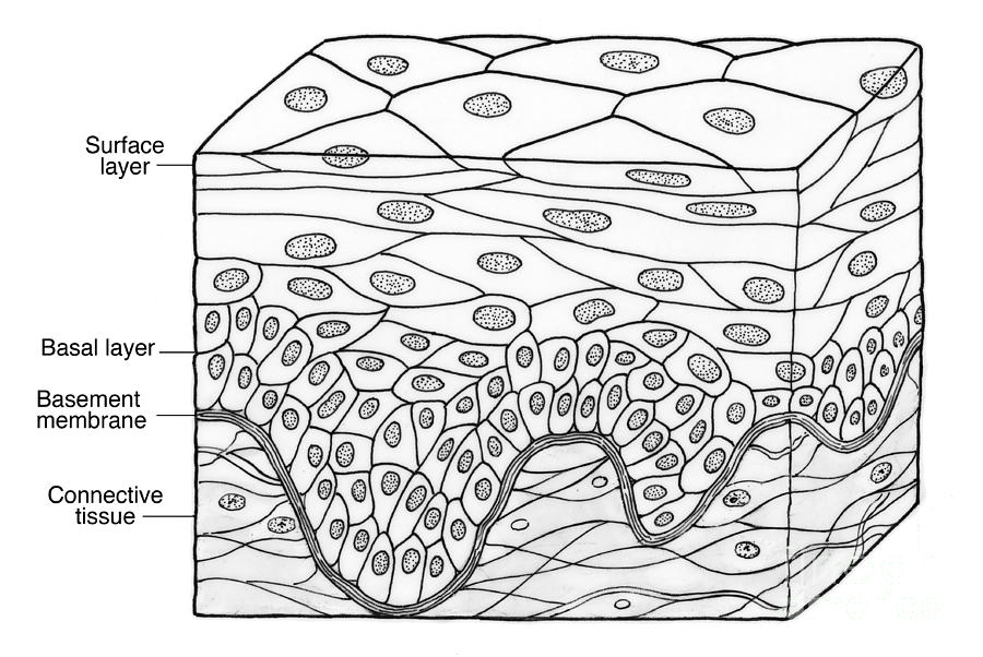
Illustration Of Stratified Squamous Photograph by Science Source Pixels
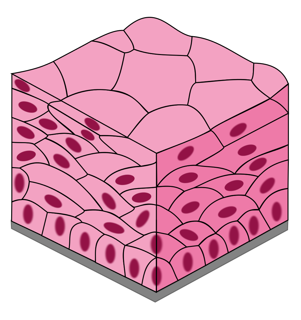
Location of simple columnar epithelium. It does not have any blood vessels within it (i.e., it is avascular). Stratified squamous epithelia are tissues formed from multiple layers of cells resting on a basement membrane, with the superficial layer (s) consisting of squamous cells. Web the epidermis is composed of keratinized, stratified squamous epithelium. Anatomy with amrutha & joseph.
Stratified squamous epithelium Function, Definition, Location, Types.
The top layer may be covered with dead cells containing keratin. Pseudostratified columnar epithelium under a. Web stratified squamous epithelium is the most common type of stratified epithelium in the human body. Web 59,228 x 34,921 pixels 7.7 gb. Only one layer is in contact with the basement membrane;

How to draw stratified squamous epithelium easy way YouTube
Web stratified squamous epithelium definition. The apical cells appear squamous, whereas the basal layer contains either columnar or cuboidal cells. O explain the composition and function of the basement membrane. Underlying cell layers can be made of cuboidal or columnar cells as well. Science source / science photo library.

Stratified Squamous Epithelium Overview, Function & Location Lesson
Web a stratified squamous epithelium consists of squamous (flattened) epithelial cells arranged in layers upon a basal membrane. This video describes how to draw stratified squamous non keratinized epithelium histology diagram. Click on to move to a specific region. The image can be changed using any combination of the following commands. Illustration of stratified squamous epithelium, showing surface layer, basal.
This Type Of Epithelium Comprises The Epidermis Of The Skin.
O classify morphological types of epithelia based on the number of cell layers, shape of apical cells, and presence of surface specializations. Stratified squamous epithelium under a microscope. Web stratified squamous epithelium is the most common type of stratified epithelium in the human body. Web here, i will provide both hand drawings and real microscope figures of stratified squamous epithelium (keratinized and nonkeratinized).
Simple Columnar Epithelium Under A Microscope.
The image can be changed using any combination of the following commands. Underlying cell layers can be made of cuboidal or columnar cells as well. It is made of four or five layers of epithelial cells, depending on its location in the body. Web 59,228 x 34,921 pixels 7.7 gb.
Skin That Has Four Layers Of Cells Is Referred To As “Thin Skin.”
Web the epidermis is composed of keratinized, stratified squamous epithelium. 4.5k views 2 years ago. Use the image slider below to learn more about the characteristics of stratified squamous epithelium. O explain the composition and function of the basement membrane.
Click On To Move To A Specific Region.
Use the image slider below to learn how to use a microscope to identify and study nonkeratinized stratified squamous epithelium lining. Location and examples of stratified squamous epithelium. Each slide is shown with additional information to its right. Web the epidermis is composed of keratinized, stratified squamous epithelium.