Thoracic Cavity Drawing
Thoracic Cavity Drawing - Right lower lobe of lung. Right upper lobe of lung. Your thoracic cavity is located in your chest. Web thoracic cavity, the second largest hollow space of the body. Web the thoracic cavity (or chest cavity) is the chamber of the body of vertebrates that is protected by the thoracic wall ( rib cage and associated skin, muscle, and fascia ). Web the thoracic cavity becomes deeper and larger, drawing in air from the atmosphere. You will want this to be large enough for you to draw more detailed structures inside. Web the thoracic cavity or the chest cavity lies between the neck and the abdomen. It’s enclosed by the bones and muscles that make up your chest wall. Right middle lobe of lung.
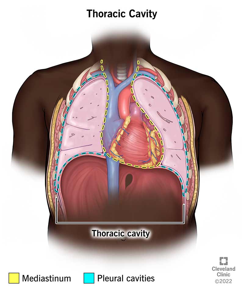
New 3d rotate and zoom. Trachea (windpipe) left lower lobe of lung. Left upper lobe of lung. Web learn more about anatomy with lecturio: The thorax is the region between the abdomen inferiorly and the root of the neck superiorly. The lungs lie either side of the mediastinum, within the thoracic cavity. For a small diagram, outline the thoracic cavity to. [1] [2] the thorax forms from the thoracic wall, its superficial structures. Your thoracic cavity is located in your chest. Web the thoracic cavity is home to many vital organs, notably the lungs/pleurae and the heart, but also includes the thymus gland and the breasts.
You will want this to be large enough for you to draw more detailed structures inside. Web thoracic cavity, the second largest hollow space of the body. For a small diagram, outline the thoracic cavity to. 152 views 1 year ago daily drawing practice. Web the thoracic cavity communicates with the neck via the superior thoracic aperture and with the abdominal cavity via the inferior thoracic aperture through anatomical spaces. The thoracic cavity (or chest cavity) is the chamber of the human body that is protected by the thoracic wall (rib cage and associated skin, muscle, and fascia), limited by the costa and the diaphragm. Web the thoracic cavity becomes deeper and larger, drawing in air from the atmosphere. Right middle lobe of lung. Left lower lobe bronchus (secondary) left main bronchus (primary) Drawn and defined [anatomy physiology] body cavities along with their organs and membranes simplified!

Thoracic Cavity by Cryssari on DeviantArt
Right upper lobe of lung. Right lower lobe of lung. It’s enclosed by the bones and muscles that make up your chest wall. Web the thoracic cavity becomes deeper and larger, drawing in air from the atmosphere. This thoracic and pulmonary anatomy tool is.
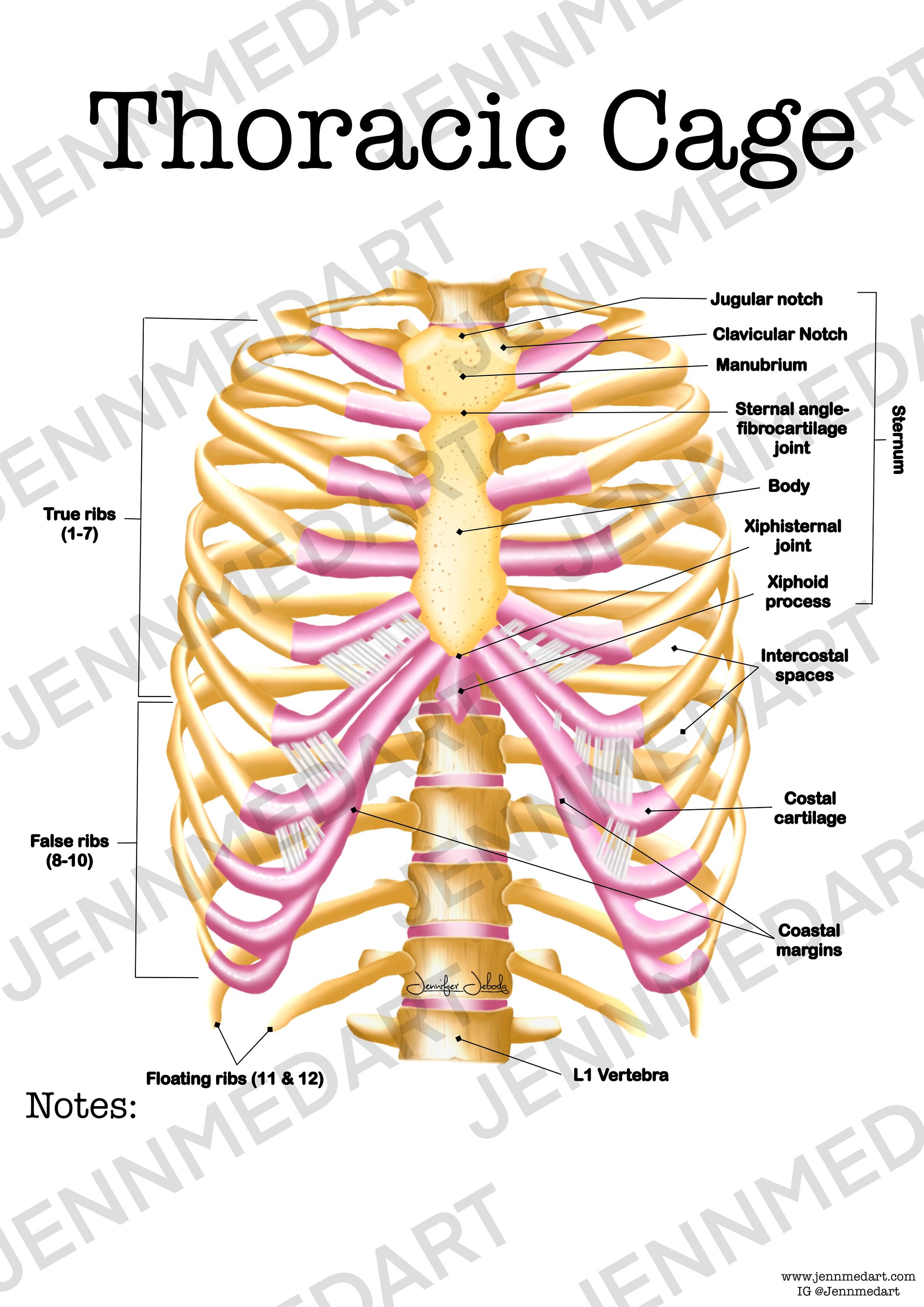
Thoracic Cage Anatomy Worksheet Single FILLED Digital Download Human
For a small diagram, outline the thoracic cavity to. [1] [2] the thorax forms from the thoracic wall, its superficial structures. This thoracic and pulmonary anatomy tool is. Web the pathway towards the lungs is provided by airways and together, these components form the respiratory system, which is located inside the thoracic or chest cavity. Web learn more about anatomy.
Thoracic Cavity Location and Function
This thoracic and pulmonary anatomy tool is. For a small diagram, outline the thoracic cavity to. Right upper lobe of lung. Web how to draw a thoracic cavity (front view) mi's creation studio. 152 views 1 year ago daily drawing practice.
Body Cavity Diagram ClipArt Best
Right upper lobe of lung. This thoracic and pulmonary anatomy tool is. During exhalation, the rib cage drops to its resting position while the diaphragm relaxes. Web the thoracic cavity or the chest cavity lies between the neck and the abdomen. Web 3d interactive modules and video tutorials on the anatomy of the thoracic cavity, including the heart, lungs, breast,.
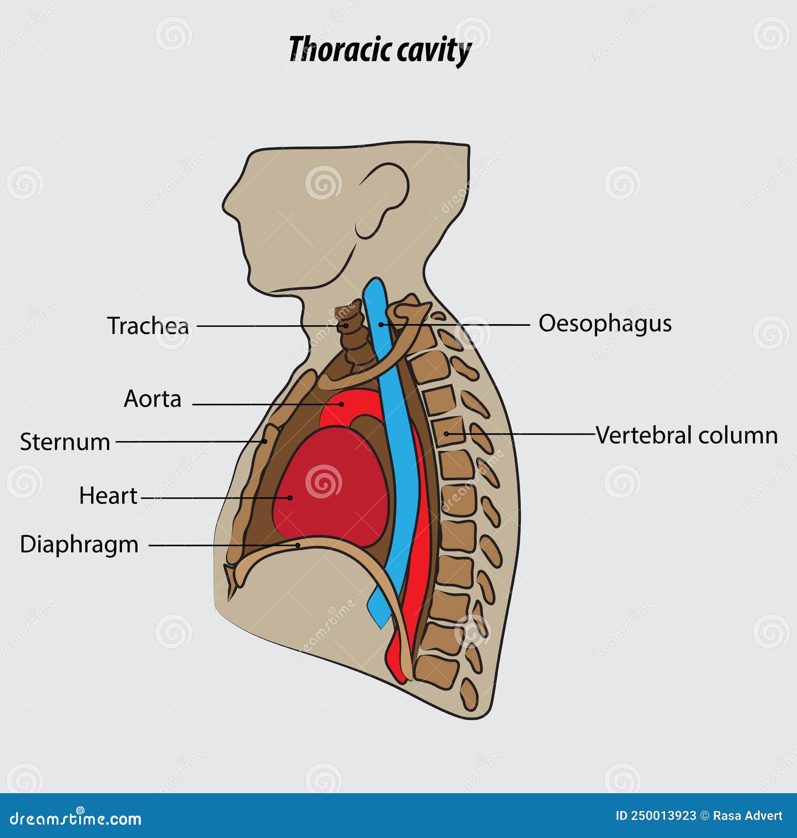
Thoracic Cavity Vector Illustration Drawing Labeled Stock Vector
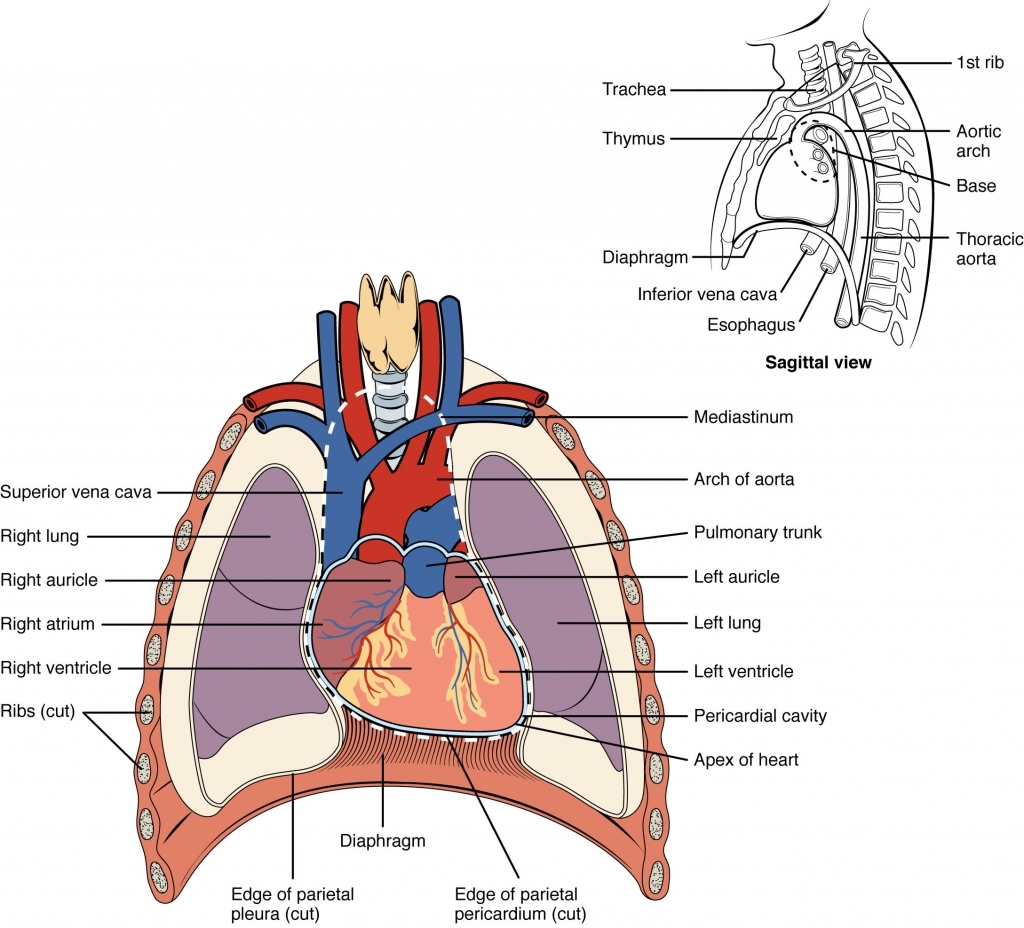
Web outline the thoracic cavity, neck, and head. The lungs lie either side of the mediastinum, within the thoracic cavity. Left lower lobe bronchus (secondary) left main bronchus (primary) New 3d rotate and zoom. Figure 19.2 shows the position of the heart within the.

Lab 2 Thoracic cavity drawing Diagram Quizlet
152 views 1 year ago daily drawing practice. Web learn more about anatomy with lecturio: For a small diagram, outline the thoracic cavity to. Web heart with blood flow. As the heart is found here, the great.
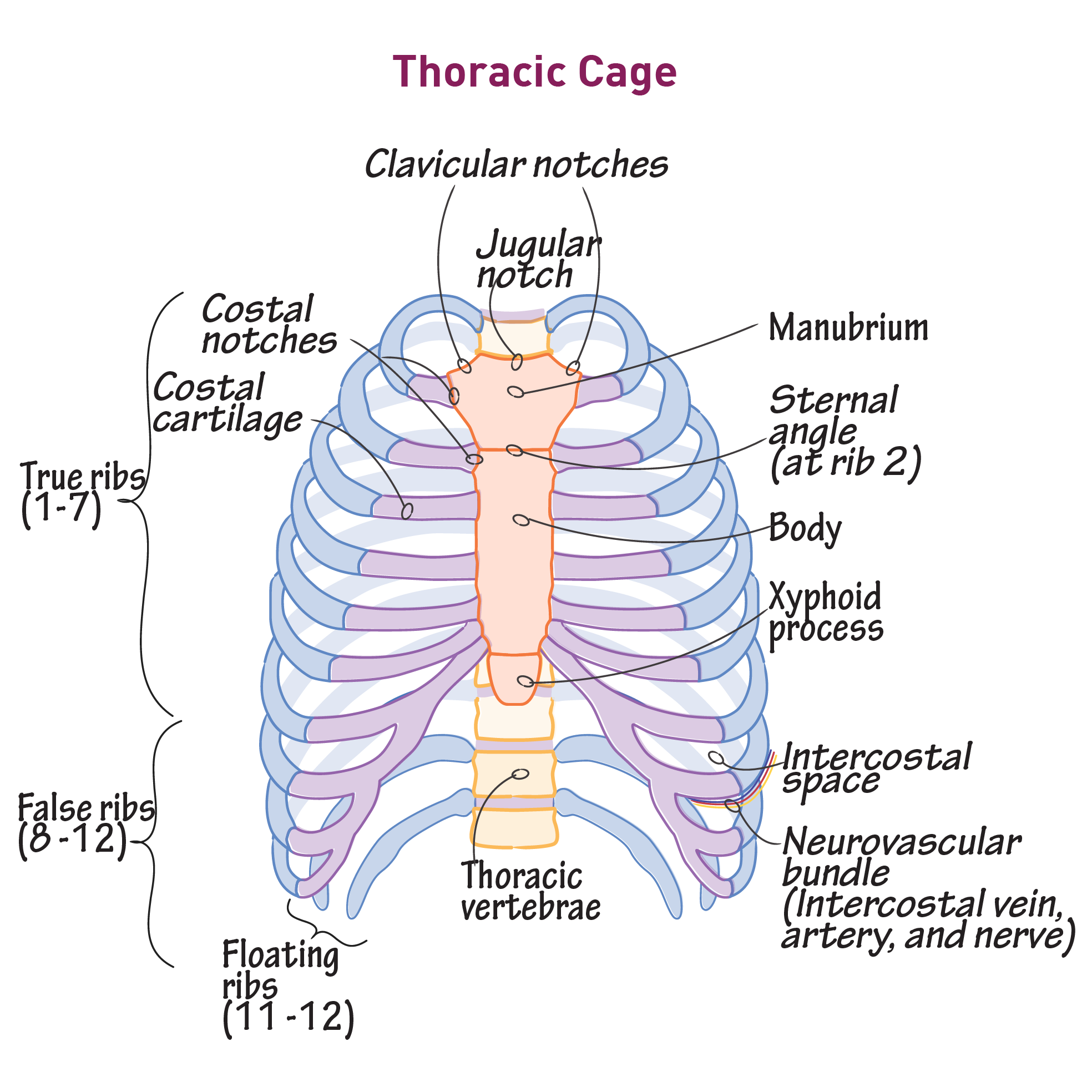
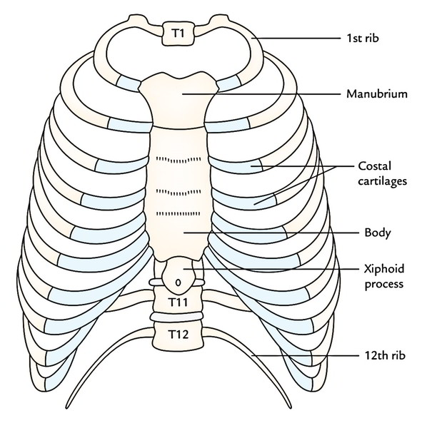
Gross Anatomy Glossary Thoracic Cage Draw It to Know It
As the heart is found here, the great. Web thoracic cavity, the second largest hollow space of the body. The thorax is the region between the abdomen inferiorly and the root of the neck superiorly. [1] [2] the thorax forms from the thoracic wall, its superficial structures. Right lower lobe of lung.
Thoracic Cage Intrinsic Muscles, Formation and Shape Earth's Lab
Web heart with blood flow. During exhalation, the rib cage drops to its resting position while the diaphragm relaxes. Web 3d interactive modules and video tutorials on the anatomy of the thoracic cavity, including the heart, lungs, breast, chest wall, and respiratory tract. Web the pathway towards the lungs is provided by airways and together, these components form the respiratory.

Human Anatomy Chest Cavity Anatomy Of Chest Bones Human Anatomy Diagram
Web the thoracic cavity (or chest cavity) is the chamber of the body of vertebrates that is protected by the thoracic wall ( rib cage and associated skin, muscle, and fascia ). Web how to draw a thoracic cavity (front view) mi's creation studio. This thoracic and pulmonary anatomy tool is. New 3d rotate and zoom. The thoracic cavity, also.

WHAT IS THORACIC CAVITY (ANATOMY) INFO HUB YouTube
Web learn more about anatomy with lecturio: Web how to draw a thoracic cavity (front view) mi's creation studio. Left upper lobe of lung. It is enclosed by the ribs, the vertebral column, and the sternum, or breastbone, and is separated from the abdominal. The thorax is the region between the abdomen inferiorly and the root of the neck superiorly.
[1] [2] The Thorax Forms From The Thoracic Wall, Its Superficial Structures.
The thorax is the region between the abdomen inferiorly and the root of the neck superiorly. Web the thoracic cavity communicates with the neck via the superior thoracic aperture and with the abdominal cavity via the inferior thoracic aperture through anatomical spaces. The thoracic cavity, also called the chest cavity, is a cavity of vertebrates bounded by the rib cage on the sides and top, and the diaphragm on the. These elements are derived from.
You Will Want This To Be Large Enough For You To Draw More Detailed Structures Inside.
Web the human heart is located within the thoracic cavity, medially between the lungs in the space known as the mediastinum. For a small diagram, outline the thoracic cavity to. It’s enclosed by the bones and muscles that make up your chest wall. Web the knowledge of the sectional anatomy of the thoracic cavity is of great importance for the understanding of respective pathologies, congenital anomalies, surgery, and radiological.
It Is Bounded By The Thoracic Wall And Extends From The Diaphragm (Below) To The Superior.
Your thoracic cavity is located in your chest. Web heart with blood flow. Web thoracic cavity, the second largest hollow space of the body. Right upper lobe of lung.
The Thoracic Wall Forms Part Of The Axial Skeleton And Is Composed Of Segmental Bone, Muscle, And Connective Tissue.
This thoracic and pulmonary anatomy tool is. Trachea (windpipe) left lower lobe of lung. Right middle lobe of lung. Left upper lobe of lung.