Ultrasound Transducer Drawing
Ultrasound Transducer Drawing - We use a commercial pzt lm transducer as our example device for calculation. Section 3 introduces the ultrasound transducer that forms the basic ultrasound transmission and reception sensor for this imaging mode. They operate at different center frequencies, have different physical dimensions, footprints, and shapes, and provide different image formats. Understand the operation of these devices. When the transducer is pressed against the skin, it directs. The transducer consists a ptz lm with d. A generator and a detector of ultrasonic waves. Understand how ultrasound images are formed. 1) physics of sound interactions in the body. Web an ultrasound transducer converts electrical energy into mechanical (sound) energy and back again, based on the piezoelectric effect.
Advantages of each transducer over the other and the technical issues for further performance enhancement are described. The operator must therefore keep in mind the orientation of the ribs wherever the transducer is being used. When the transducer is pressed against the skin, it directs. The ultrasound transducer generates ultrasound (ultrasonic) waves. 3) relative strengths and weaknesses of different transducers including various aspects of resolution. Physics and safety, understand the following: Ultrasound waves are emitted rapidly from the transducer. The ultrasound transducer & piezoelectric crystals. Understand how ultrasound images are formed. Web since it is a very commonly used ultrasonic transducer perhaps someone knows how to calculate the maximum current draw at a specific frequency and voltage, and/or a better datasheet or information or a close enough estimation of what current it will draw or what unit the value is in.
Web based on the wave equation and frequency equation, an analytical model of the ultrasonic vibration squeeze transducer is established, and the structural dimensi design and simulation analysis of ultrasonic extrusion transducer | ieee conference publication |. Location and orientation of the ultrasound transducer for echocardiography, over the anterior intercostal spaces. When the transducer is pressed against the skin, it directs. Web the types of transducers are classified according to the dimensions of ultrasound images: 33= 400pm/v and 800 m thickness. The operator must therefore keep in mind the orientation of the ribs wherever the transducer is being used. The diagrams show the sound fields of an unfocused and a focusing ultrasonic transducer in water, plainly at differing energy levels. Explain how transducers can electronically focus and steer the ultrasound beam. Section 3 introduces the ultrasound transducer that forms the basic ultrasound transmission and reception sensor for this imaging mode. Web a pdms layer and coupling gel are used between the transducer and the skin to achieve near perfect transmission of the ultrasonic signal.
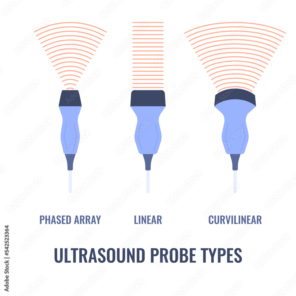
Ultrasound probe types diagram. Linear, curvilinear and phased array
The transducer consists a ptz lm with d. Linear array transducers produce rectangular images with width of the image determined by the physical width of the transducer face. 33= 400pm/v and 800 m thickness. Web overcome this effect is to rotate the transducer so that it lies entirely within the space between the ribs. There are different types of transducers.
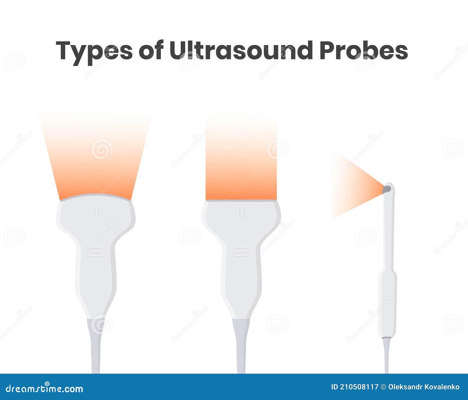
Types of Ultrasound Tranducers Convex, Linear and Encocavitary Probes
Web an ultrasound transducer converts electrical energy into mechanical (sound) energy and back again, based on the piezoelectric effect. Web operating an ultrasound unit. Web the illustration shows a schematic drawing of wave length, pressure and amplitude. Web in the past few decades, medical ultrasound array design has been an active research area with challenging technical requirements that continually seek.

Ultrasonics Transducers Piezoelectric Hardware CTG Technical Blog
Vector transducers provide slightly wider near field of view than do sector transducers. Web download scientific diagram | ultrasonic transducer (after wells 1977): Location and orientation of the ultrasound transducer for echocardiography, over the anterior intercostal spaces. Selecting the image mode, gain, and focus; The transducer is held with one hand and its position and angle are adjusted to send.
Piezoelectric Transducer Simulation with OnScale Ultrasonic Sensor
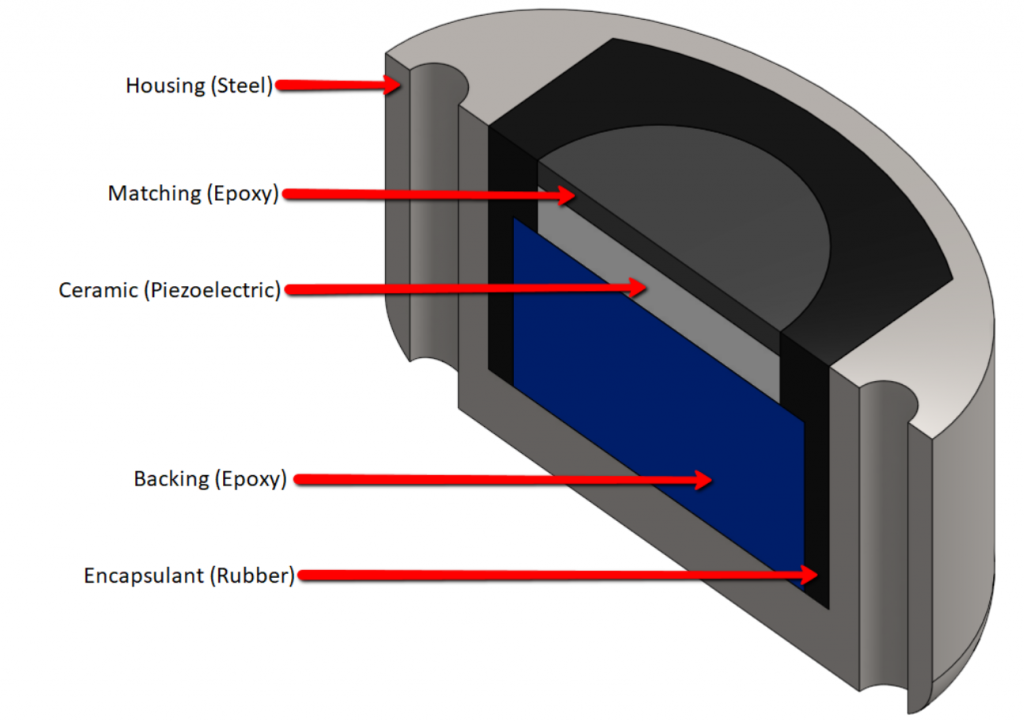
Explain how transducers can electronically focus and steer the ultrasound beam. A transducer consists of five main components: Location and orientation of the ultrasound transducer for echocardiography, over the anterior intercostal spaces. The transducer is held with one hand and its position and angle are adjusted to send ultrasound waves through structures to be visualized. Transducer materials and probe construction.
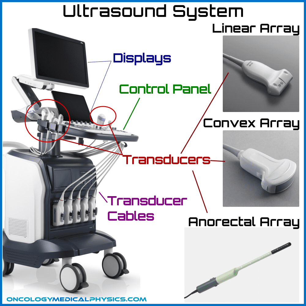
Ultrasound Design and Operation Oncology Medical Physics
Many types of medical ultrasound transducers are used in clinical practice. Web download scientific diagram | ultrasonic transducer (after wells 1977): Explain how transducers can electronically focus and steer the ultrasound beam. The electrical signals are transmitted to the target by these transducers and after the signal reaches the object, it returns to the transducer. Web overcome this effect is.

The ultrasound transducer ECG & ECHO
It converts mechanical energy into electrical energy and vice versa. Web 25 february 2021 9503. In this section, the components that a modern ultrasound system are based on are provided along with a brief description of ultrasound properties applicable to imaging. Web an ultrasound transducer functions as both: Linear array transducers produce rectangular images with width of the image determined.

Phasedarray transducers. A. A typical ultrasound transducer for
Selection of transducer and preset; The ultrasound transducer generates ultrasound (ultrasonic) waves. Many types of medical ultrasound transducers are used in clinical practice. This transducer tests the distance of the object in this method, not the amplitude of the signal. Web overcome this effect is to rotate the transducer so that it lies entirely within the space between the ribs.
Ultrasound Drawing at GetDrawings Free download
They operate at different center frequencies, have different physical dimensions, footprints, and shapes, and provide different image formats. Understand the operation of these devices. Web a pdms layer and coupling gel are used between the transducer and the skin to achieve near perfect transmission of the ultrasonic signal. Web the types of transducers are classified according to the dimensions of.
Ultrasound Drawing at GetDrawings Free download
Web 25 february 2021 9503. Understand how ultrasound images are formed. Web an ultrasound transducer functions as both: Section 3 introduces the ultrasound transducer that forms the basic ultrasound transmission and reception sensor for this imaging mode. Advantages of each transducer over the other and the technical issues for further performance enhancement are described.

An illustration of ultrasound transducers in an ultrasound system
A transducer consists of five main components: Web operating an ultrasound unit. Section 3 introduces the ultrasound transducer that forms the basic ultrasound transmission and reception sensor for this imaging mode. Transducer materials and probe construction. 33= 400pm/v and 800 m thickness.
Understand How Ultrasound Images Are Formed.
The resulting design achieves high e ciency and can handle transducer impedance variations by adjusting two capacitances in the matching network. When the transducer is pressed against the skin, it directs. Transducer materials and probe construction. Web operating an ultrasound unit.
Selection Of Transducer And Preset;
For most humans audible sound ranges between 20 hz and 20,000 hz (20 khz) ultrasound refers to any sound waves with frequencies greater than 20khz. Web the beam pattern of a transducer can be determined by the active transducer area and shape, the ultrasound wavelength, and the sound velocity of the propagation medium. The ultrasound transducer & piezoelectric crystals. A transducer consists of five main components:
Linear Array Transducers Produce Rectangular Images With Width Of The Image Determined By The Physical Width Of The Transducer Face.
Web the illustration shows a schematic drawing of wave length, pressure and amplitude. Web 25 february 2021 9503. The operator must therefore keep in mind the orientation of the ribs wherever the transducer is being used. Selecting the image mode, gain, and focus;
Web A Pdms Layer And Coupling Gel Are Used Between The Transducer And The Skin To Achieve Near Perfect Transmission Of The Ultrasonic Signal.
Understand the operation of these devices. It converts mechanical energy into electrical energy and vice versa. Section 3 introduces the ultrasound transducer that forms the basic ultrasound transmission and reception sensor for this imaging mode. Web since it is a very commonly used ultrasonic transducer perhaps someone knows how to calculate the maximum current draw at a specific frequency and voltage, and/or a better datasheet or information or a close enough estimation of what current it will draw or what unit the value is in.